Why Treating Complex Melasma Requires a Team Versed in Dermatopathology
Every year, thousands of melasma patients cycle through dermatology clinics, medical spas, and laser centers, receiving treatments that target what is visible on the surface — the brown or grey-brown patches — while ignoring the deeper pathological processes driving the condition. The result is a frustrating loop: temporary fading, aggressive rebound, worsening hyperpigmentation, and progressive skin sensitization. The missing piece is not a better laser or a stronger peel. It is a fundamental understanding of dermatopathology — the microscopic disease processes occurring within the skin itself.
At Liusmed Clinic, we believe that melasma treatment without dermatopathological reasoning is like prescribing antibiotics without understanding microbiology. This article explains why complex melasma demands a team that thinks at the tissue level, and how that approach changes outcomes.
Table of Contents
- Beyond the Surface: What Dermatopathology Reveals About Melasma
- The Four Pillars of Melasma Pathology
- Why Single-Specialty Clinics Often Fail
- What a Dermatopathology-Informed Team Looks Like
- Comparing Treatment Paradigms: Surface vs. Tissue-Level
- How Liusmed Clinic Applies This Philosophy
- Frequently Asked Questions
- About the Author
- Disclaimer
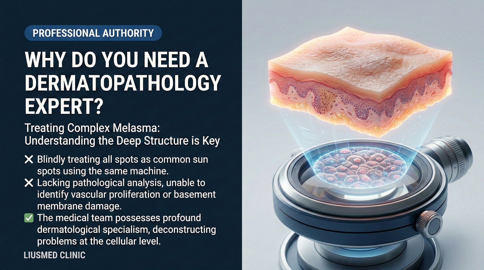
Beyond the Surface: What Dermatopathology Reveals About Melasma {#beyond-the-surface}
When a pathologist examines a melasma biopsy under the microscope, they do not simply see excess melanin. They see a constellation of interrelated abnormalities that explain why this condition is so resistant to conventional treatments.
The epidermis shows increased melanocyte activity, but that is only the beginning. The dermal-epidermal junction — the basement membrane zone — frequently shows structural disruption. Melanin granules that should remain in the epidermis have dropped into the dermis, where macrophages engulf them and form stubborn deposits called melanophages. These dermal melanin deposits are largely unresponsive to topical lightening agents because those agents cannot penetrate deeply enough to reach them.
Below the basement membrane, the dermis reveals another critical finding: increased vascularity. The blood vessels in melasma-affected skin are more numerous and more dilated than in normal skin. This is not a coincidence. Vascular endothelial growth factor (VEGF) is elevated in melasma lesions, and the increased blood supply creates a pro-inflammatory, pro-melanogenic microenvironment that continuously stimulates melanocytes from below.
Additionally, solar elastosis — the degradation of dermal collagen and elastic fibers from chronic UV exposure — is nearly always present. This damaged dermal matrix provides a permissive environment for ongoing pigmentary dysfunction.
Understanding these layers of pathology is what separates a dermatopathology-informed approach from a purely cosmetic one.
The Four Pillars of Melasma Pathology {#four-pillars}
Effective melasma treatment must address four distinct but interconnected pathological processes:
1. Melanocyte Hyperactivity in the Epidermis
Melanocytes in melasma skin are not more numerous — they are hyperactive. They produce melanin at an accelerated rate in response to UV radiation, hormonal signals, heat, and inflammatory cytokines. Any treatment that ignores these triggers while trying to destroy melanin is fighting a battle it cannot win.
2. Basement Membrane Disruption
The basement membrane acts as a gatekeeper, preventing melanin from falling into the dermis. In melasma, this barrier is compromised. Aggressive treatments — including overly frequent laser sessions and deep chemical peels — can worsen this disruption, paradoxically making the condition harder to treat over time.
3. Dermal Vascular Proliferation
The increased vascular network in melasma skin delivers more nutrients, more inflammatory mediators, and more hormonal signals to the melanocytes above. Treatments that address vascularity — such as tranexamic acid, which has anti-angiogenic properties — target this pillar directly.
4. Chronic Low-Grade Inflammation
Mast cells, inflammatory cytokines, and oxidative stress markers are elevated in melasma tissue. This chronic inflammatory milieu keeps melanocytes in a perpetual state of activation. Any treatment that induces significant inflammation (such as aggressive lasers) risks worsening the very condition it aims to treat.
Why Single-Specialty Clinics Often Fail {#single-specialty-failure}
Most melasma patients seek treatment at one of three types of facilities, each with inherent limitations:
Dermatology clinics often rely on topical agents (hydroquinone, retinoids, azelaic acid) and procedural interventions (chemical peels, low-fluence lasers). While these address epidermal melanin, they rarely account for dermal vascularity or basement membrane integrity in a systematic way.
Medical spas and laser centers tend to focus on device-based treatments. The emphasis is on technology — picosecond lasers, Q-switched (Q-switched laser) lasers, intense pulsed light — without sufficient understanding of when these devices help and when they cause harm. The risk of post-inflammatory hyperpigmentation (PIH) and rebound melasma is significant, particularly in patients with darker skin tones or Fitzpatrick (Fitzpatrick Skin Type) types III-V.
Aesthetic medicine practices may offer injectable treatments like tranexamic acid or PRP (Platelet-Rich Plasma — concentrate of your own blood platelets rich in growth factors), but without the dermatological training to properly diagnose the type and depth of pigmentation, or to recognize contraindications.
The fundamental problem is that complex melasma requires knowledge that spans dermatology, pathology, vascular biology, and regenerative medicine. No single specialty covers all of these domains adequately.
What a Dermatopathology-Informed Team Looks Like {#informed-team}
A team capable of treating complex melasma should include the following competencies:
Diagnostic acuity. The ability to differentiate melasma from other pigmentary disorders — including post-inflammatory hyperpigmentation, lichen planus pigmentosus, exogenous ochronosis, and drug-induced pigmentation — is essential. Misdiagnosis leads to inappropriate treatment and wasted time.
Histological reasoning. Even without performing biopsies on every patient, the treating physician must think in histological terms. Is this primarily epidermal or dermal pigment? Is the basement membrane intact? Is there significant vascularity? These questions guide every treatment decision.
Vascular assessment. Understanding the role of dermal vascularity in melasma maintenance, and having tools and techniques to address it, is critical. This includes both pharmacological approaches (tranexamic acid, anti-inflammatory agents) and procedural approaches (vascular-targeting devices when appropriate).
Regenerative medicine expertise. Repairing the basement membrane, restoring dermal collagen architecture, and modulating inflammation all fall within the domain of regenerative medicine. Techniques like PRP therapy, growth factor delivery, and controlled wound-healing protocols can rebuild the tissue environment that conventional treatments ignore.
Comparing Treatment Paradigms: Surface vs. Tissue-Level {#comparing-paradigms}
| Dimension | Surface-Level Approach | Dermatopathology-Informed Approach |
|---|---|---|
| Diagnostic focus | Visual appearance of pigmentation | Tissue-level pathology (epidermal, dermal, vascular) |
| Primary tools | Lasers, peels, topical lightening | Multi-modal: topical, injectable, regenerative, vascular |
| Basement membrane | Not specifically assessed | Central treatment target |
| Vascularity | Ignored or incidentally treated | Directly addressed with anti-angiogenic strategies |
| Inflammation management | Reactive (treat PIH after it occurs) | Proactive (minimize inflammatory triggers) |
| Treatment timeline | Short-term (4-8 sessions) | Long-term (phased repair over 6-12+ months) |
| Recurrence strategy | Repeat same treatment | Maintenance protocol addressing root causes |
| Typical outcome | Temporary improvement, frequent rebound | Sustained improvement with lower recurrence |
How Liusmed Clinic Applies This Philosophy {#liusmed-approach}
At Liusmed Clinic, our Melasma Injection Treatment protocol is built on dermatopathological principles. Rather than defaulting to a single device or product, we construct individualized treatment plans based on the specific pathological profile of each patient's melasma.
Phase 1: Comprehensive Assessment. We evaluate the depth and distribution of pigmentation, the degree of vascular involvement, the integrity of the skin barrier, and the patient's treatment history. Patients who have undergone extensive prior laser treatment often present with compromised basement membranes and sensitized skin, requiring a period of stabilization before active treatment.
Phase 2: Tissue Environment Repair. Before aggressively targeting pigment, we focus on restoring the conditions necessary for healthy melanocyte function. This includes basement membrane support, inflammation reduction, and vascular modulation. PRP therapy and targeted injectable protocols play a central role in this phase.
Phase 3: Controlled Pigment Reduction. Only after the tissue environment is stabilized do we employ pigment-targeting interventions — and even then, with conservative parameters designed to minimize inflammatory rebound. The goal is gradual, sustained improvement rather than dramatic but temporary clearing.
Phase 4: Maintenance and Monitoring. Complex melasma is a chronic condition. We design maintenance protocols that patients can sustain long-term, including topical regimens, periodic in-clinic treatments, and lifestyle modifications (UV protection, heat avoidance, hormonal management where applicable).
This phased, tissue-level approach is why patients who have failed multiple treatments elsewhere often find sustained improvement at Liusmed Clinic.
Frequently Asked Questions
Q1: What is dermatopathology and why does it matter for melasma?
Dermatopathology is the study of skin diseases at the microscopic tissue level. For melasma, it matters because the condition involves changes not just in melanin production but in basement membrane integrity, dermal vascularity, and chronic inflammation. A dermatopathology-informed approach addresses all of these factors rather than just targeting visible pigmentation.
Q2: Can melasma be cured, or only managed?
Current medical understanding classifies melasma as a chronic, relapsing condition. While significant and sustained improvement is achievable — especially with a tissue-level treatment approach — most patients benefit from ongoing maintenance protocols. The goal is long-term control with minimal recurrence rather than a one-time cure.
Q3: Why did my melasma get worse after laser treatment?
Aggressive laser treatment can worsen melasma through several mechanisms: inducing post-inflammatory hyperpigmentation, further disrupting an already compromised basement membrane, and triggering inflammatory cascades that stimulate melanocyte activity. This is particularly common when treatment parameters are not carefully calibrated for melasma-specific pathology.
Q4: How is a dermatopathology-informed approach different from what my dermatologist offers?
Many dermatologists provide excellent melasma care. The distinction is not about specialty credentials per se, but about treatment philosophy. A dermatopathology-informed approach systematically evaluates and addresses all four pillars of melasma pathology (melanocyte activity, basement membrane integrity, vascularity, and inflammation) rather than focusing primarily on pigment reduction.
Q5: What role does PRP play in treating complex melasma?
PRP (platelet-rich plasma) contains growth factors that support tissue repair, including basement membrane restoration, collagen remodeling, and anti-inflammatory modulation. In the context of melasma, PRP can help rebuild the tissue environment that has been disrupted by the disease process and by prior aggressive treatments. Learn more about our Melasma Injection Treatment approach.
Q6: How long does a tissue-level melasma treatment plan typically take?
Complex melasma treatment typically unfolds over 6 to 12 months or longer, depending on the severity of the condition and the patient's treatment history. Patients with extensive prior laser damage or deeply entrenched dermal pigmentation may require longer timelines. The phased approach — stabilization, repair, controlled reduction, maintenance — ensures that each stage builds on the progress of the previous one.
About the Author
Dr. Ta-Ju Liu is the founder of Liusmed Clinic, a specialized practice integrating regenerative medicine with minimal incision surgical techniques. With dual expertise in dermatological science and tissue repair, Dr. Liu has developed treatment protocols for complex pigmentary disorders — including refractory melasma — that prioritize tissue-level healing over surface-level cosmetic correction. His approach reflects a commitment to understanding and addressing the root pathological mechanisms behind conditions that resist conventional treatment.
Disclaimer
This article is provided for educational and informational purposes only and does not constitute medical advice, diagnosis, or treatment. Individual conditions vary, and treatment outcomes depend on numerous patient-specific factors. Always consult a qualified healthcare professional for personalized medical guidance. The information presented here reflects the clinical perspective of Liusmed Clinic and should not be taken as a guarantee of treatment results.

Specialties
Credentials
- Kaohsiung Medical University, School of Medicine
- Attending Physician, Dermatology, Kaohsiung Chang Gung Memorial Hospital
- Attending Physician, Aesthetic Center, Kaohsiung Chang Gung Memorial Hospital
- Visiting Physician, Dermatology, Xiamen Chang Gung Hospital
- Visiting Physician, Aesthetic Center, Xiamen Chang Gung Hospital
"For every surgery, I strive to achieve a good outcome through a small incision and refined technique. Minimally invasive surgery is not just a technique — it's a commitment of respect to every patient."
Recovery after any procedure needs peer support too
Want to learn more?
Schedule a consultation for professional evaluation and advice