
Fat Graft Complication Repair Authority Guide
From Facial Overfilling and Calcified Lumps to Under-Eye GranulomasThe Ultimate Rescue with Micro-Pinhole Extraction
Although fat grafting uses your own tissue, complications can be equally challenging — facial overfilling syndrome (FOS), calcified lumps, and under-eye fat granulomas (caterpillar-like bumps) are problems that traditional liposuction or steroid injections often cannot resolve. Dr. Liu at Liusmed Clinic specializes in ultrasound-guided "Micro-Pinhole Extraction," precisely removing fibrotic fat masses through just 1-2mm micro-incisions, restoring natural contours without major surgery.
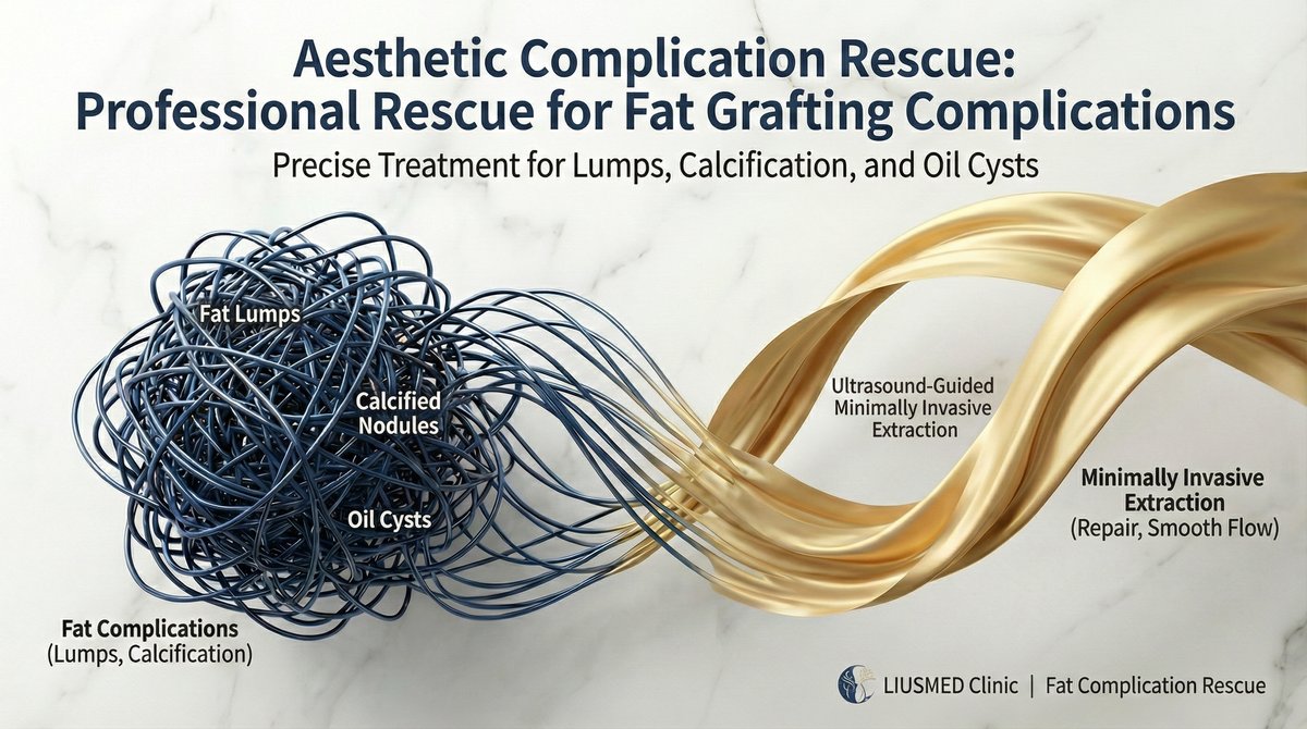
Table of Contents
Are You Experiencing These Issues?
Fat graft complications are more common than many people realize. Here are the most frequent issues:
Common Fat Graft Complications
These problems will not resolve on their own over time, but they can be safely addressed with the right minimally invasive technique.
Pathological Mechanisms: Why Do Fat Grafts Go Wrong?
Although fat grafts use "your own" tissue, not all transplanted fat cells survive. Understanding the pathology is key to effective treatment.
Central Necrosis → Fibrous Encapsulation → Calcification
The Fate of Fat Masses
When the radius of a single-point fat injection exceeds 2 mm, the core of the fat bolus cannot receive sufficient oxygen diffusion or neovascular supply within the first 48 hours, resulting in central necrosis. Necrotic fat releases free fatty acids, triggering sterile inflammation — the immune system dispatches macrophages to form a dense, rigid fibrous capsule wall. This is why the lump feels as hard as a "stone." Over time, calcium deposits accumulate within the necrotic core, forming permanent calcified nodules that no lipolytic injection can dissolve.
FOS: Compartment Destruction & Expression Restriction
Chain Reaction of Excess Fat
In pursuit of volume, excessive fat is injected into both the superficial and deep fat compartments, destroying the face's precisely organized "retaining ligament" architecture. Fat overflows into layers where it shouldn't exist, compressing the mimetic muscles — when fat is inadvertently injected into the muscular layer or accumulates on top of expression muscles, muscular contraction meets mechanical resistance, producing the characteristic "plastic" appearance with every smile. More filling leads to more swelling, which leads to looking older — that is the essence of FOS.
Periorbital: Granuloma & Permanent Edema
The Truth Behind Under-Eye "Caterpillars"
The under-eye skin is approximately only 0.5 mm thick — the thinnest on the entire face. Necrotic fat cells after grafting trigger granulomatous reactions, forming visible "caterpillar"-like strip or granular bumps under directional lighting. Worse still, excessive fat masses compress the delicate periorbital lymphatic network, obstructing lymphatic drainage and causing chronic, non-resolving malar edema. This swelling will not resolve over time — it can only improve by physically removing the offending tissue.
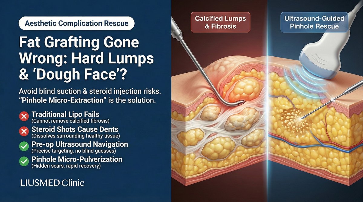
Why Traditional Methods Fail: The Dead Ends of Conventional Repair
Repairing fat graft complications is far more difficult than conventional fillers because fat integrates tightly with native tissue and fibrosis is more severe. Critically, unlike hyaluronic acid fillers, fat has no dissolver — once calcified, no enzyme can break it down. This is why most doctors say "nothing can be done." Here are the limitations of traditional methods:
Traditional Liposuction
Liposuction cannulas are designed to aspirate soft, mobile fat. Against dense fibrotic lumps, the cannula simply glides over the surface like sliding across a stone — unable to gain purchase. Forceful manipulation only damages the surrounding healthy tissue, creating surface irregularities and making the problem worse.
Steroid Injections
Killing one hundred enemies, wounding three thousand allies. Steroids cannot selectively target the lump — the typical outcome: the hard core persists while surrounding healthy fat atrophies, creating unsightly "volcanic crater" depressions. Completely ineffective against calcified lumps, and actually increases the complexity of subsequent repair.
Laser Lipolysis
Dissolving calcified deposits requires elevated laser energy. In thin-skinned areas like the forehead and periorbital region, this substantially increases the risk of thermal skin burns and nerve heat injury. Laser heat also cannot precisely distinguish fibrotic tissue from normal tissue, easily causing irreversible collateral damage.
Open Excision
Although the most thorough approach, making a 2-3 cm incision on the face and accepting a permanent scar is simply unacceptable for patients seeking aesthetic improvement. Extensive tissue dissection also risks nerve damage and tissue collapse, with significantly prolonged recovery.
Core Technology: Ultrasound-Guided Micro-Pinhole Extraction
Four-Step Precision Repair: From Localization to Aesthetic Reshaping
High-Frequency Ultrasound Guidance
Visualized Surgery
We refuse to operate blindly. Using high-resolution soft-tissue ultrasound, we perform a full-face tomographic scan before the procedure. This precisely maps the depth, nature (liquid oil cyst vs. calcified lump), and surrounding vascularity of each deposit. This "navigation map" is the cornerstone of surgical success — giving the surgeon a complete subcutaneous roadmap before any incision.
Precision Micro-Dissection
Separating Lesion from Normal Tissue
When working in high-risk zones such as the forehead (supratrochlear nerve territory) and periorbital region, safety is the paramount principle. Through a mere 1-2mm pinhole entry, specialized micro-instruments gently dissect the lump away from surrounding neurovascular structures, creating a safe buffer zone and preserving healthy tissue before proceeding.
Pinhole Micro-Fragmentation & Extraction
Breaking Down and Removing Piece by Piece
This is the technical heart of the procedure. We abandon the crude liposuction cannula in favor of microsurgical energy devices. Hardened calcified deposits are pulverized "in situ" beneath the skin — breaking the large "stone" into fine "sand" particles before precise aspiration, completely removing stubborn calcifications that even laser cannot eliminate. The entire process requires only a 1-2mm pinhole incision.
Subtractive Aesthetics & Tissue Repositioning
Not Just Removal, But Restoration
To address skin laxity from FOS correction, we sculpt the subcutaneous space and apply "skin re-adherence techniques," allowing the skin to re-adhere naturally to the deeper tissue framework, restoring the face's original bony contours. The core philosophy of "subtractive aesthetics": removing what's excess requires more skill than adding more — it's not just removal, but restoration.
The Liusmed Standard: Clean, Even, and Precisely Measured Removal
Many patients have already had a removal elsewhere before coming to Liusmed — but "having had it removed" does not mean it was removed cleanly, and certainly not evenly. The most common situation we see is a surface left bumpy after removal, sometimes with a brand-new hollow. For us, whether something can be extracted was never the point. What truly matters is whether what remains is clean, even, and precise enough to be "neither too much nor too little."
Clean Removal
We remove the stubborn mass thoroughly, leaving no residual fragments that would let the problem recur and become harder to treat later.
Even Surface
After extraction the surface stays smooth — no new craters or wavy irregularities. The most common failure is over-removal that creates a fresh hollow; avoiding it takes restraint and feel, not aggressiveness.
Precise, Not Excessive (Fat Grafting in Reverse)
Before we begin, we map out the raised areas across the whole face and account for how your muscles move the fat when you smile or talk — so that once swelling settles, exactly the right amount is removed, no more and no less. It is precision fat grafting performed in reverse.
This feel for evenness and precision comes from Dr. Ta-Ju Liu's years of underarm rotary-blade surgery, liposuction-and-grafting, and liposuction revision — drawing on the same judgment of facial fat layers and dynamics.
At follow-up one to two months after the procedure, most patients give positive feedback on how even and natural their contours look.
FAQ
Can calcified fat graft lumps from years ago still be treated?
Yes. Regardless of how much time has passed, as long as the lump still exists and causes problems, we can precisely treat it with ultrasound-guided minimally invasive methods. Even stone-hard calcified lumps can be safely removed through our pinhole micro-fragmentation extraction technique.
Will my face look sunken after fat removal?
This is the most common concern, but the answer is actually the opposite. Overfilling or excess fat makes you "look older." After precisely removing the excess and displaced fat, facial contours actually return to a younger, more natural state. We apply "subtractive aesthetics" philosophy combined with tissue repositioning techniques to ensure smooth, natural post-operative contours.
Can under-eye fat granulomas (caterpillars) be treated?
Yes. Under-eye fat granulomas are among the trickiest complications of fat grafting. We use ultrasound to precisely locate the granuloma's layer and extent, then carefully extract through a micro-pinhole approach while avoiding the delicate periorbital neurovascular network. Recovery takes about 1-2 weeks, restoring smooth, natural under-eye contours.
Is general anesthesia required? How long is recovery?
Most cases require only local anesthesia with no hospitalization needed. The wound is just 1-2mm pinhole-sized. There may be mild swelling and bruising post-op, typically recovering for normal social activities within 1-2 weeks. Recovery time is significantly shorter compared to traditional open surgery.
Why aren't steroids recommended for fat graft lumps?
Steroids indiscriminately dissolve normal fat, creating "crater" depressions that are even harder to repair. Moreover, steroids are completely ineffective against calcified or fibrotic masses. Clinically, patients who received steroid injections often develop more severe surface irregularities, actually increasing the complexity of subsequent repair.
How is pricing determined?
Fat graft repair is a highly customized medical procedure. Cost depends on the number of fat masses, their depth and location, degree of fibrosis/calcification, and severity. Pricing requires the doctor's hands-on examination and ultrasound assessment. We insist on providing only necessary treatment without wasting patients' money.
Repair Process
Precise Assessment × Micro Repair × Complete Follow-up
Consultation
Understanding your situation, fat graft history, and expectations with hands-on examination and ultrasound assessment
Treatment Planning
Developing personalized plan and pricing based on assessment
Micro Repair
Precise ultrasound-guided minimally invasive repair, wound only 1-2mm
Follow-up Care
7-14 day post-op follow-up to ensure smooth recovery

Dr. Ta-Ju Liu
Director, Liusmed Clinic
Specialties
Credentials
- over 15 years clinical minimally invasive surgery experience
- One of the few specialists in Taiwan focused on "fat graft complication repair"
- Developed ultrasound-guided micro fat repair techniques
"Real medicine is not about adding icing to the cake, but being willing to stand by patients when problems arise, becoming the last stop that can solve their suffering."
Online Initial Assessment Tool
60-Second Filler Over-Filling Self-Check
8 quick yes/no questions (stiffness, puffiness, friends' feedback, lumps, asymmetry, expression range, sessions, overall fullness). Weighted score determines a 4-tier risk level. The result page lets you one-tap pre-fill the consultation form or send your result to the clinic via LINE. No appointment needed.
Posted in the forum? We can help expedite your appointment.
Standard booking takes 3+ months. If you post your case in the FillerRescue forum first and then reach out via the button below with the required info, we’ll watch for earlier slots and help arrange your appointment as soon as possible.
You're Not Alone in This
The road to post-procedure repair needs more than medical expertise — whether it is filler, fat grafts, scars, energy-device damage, threads, post-surgical concerns, complex skin disease, or even surgical failure and recurrence, it takes reliable information and peer support. This is a community for aesthetic post-procedure repair, offering doctor-led education and patient-to-patient connection.
Doctor Community
fillerrevision.com
Educational content posted by Dr. Liu — daily updates, case analyses, and knowledge sharing. Get first-hand information from a repair specialist.
FillerRescuePatient Forum
Where patients come first. Always.
A nonprofit community forum where patients lead the conversation. Q&A, shared experiences, recovery journeys, and an SOS emergency feature. Post anonymously and receive responses reviewed by a medical expert panel.
From pillow face to lumps to under-eye caterpillars — micro-pinhole extraction repair
Fat is "your own tissue" — but complications are equally tricky; ultrasound-classified + 1-2mm pinhole extraction
Your Fat Graft Repair Includes
Ultrasound-guided classification · live fat vs. necrotic fibrous masses
Remove only what must come out · preserve functional fat · avoid contour defects
1-2mm pinhole · no scalpel, no sutures
Handles central necrosis · fibrous encapsulation · calcified masses
Addresses all 3 pathological stages of fat graft failure · no open surgery
LIUSMED integrated post-op repair system included
※ Click any chip to view full scope and exclusion terms
Want to know which path fits your situation? Either way works — pick whichever feels easier.
Other Repair Services
Real medicine means standing by patients when problems arise.
We hope Liusmed can be your "rescue station."
Don't Let Failed Fat Grafts Hold You Back
Stop suffering alone, give yourself a chance to start fresh
Perhaps we are the ones who can help solve your problem
Book Fat Graft Revision Consultation