Facial Spider Veins: Can Skincare Really Remove Them?
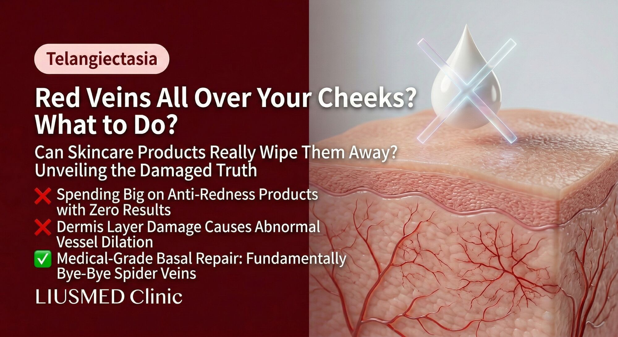
Those tiny red or purple branching lines on your cheeks and nose are not just cosmetic imperfections. Facial telangiectasia, commonly called spider veins, represent structurally altered blood vessels that have permanently dilated and lost the ability to constrict back to normal size. Despite the bold claims on countless skincare product labels, no topical cream, serum, or essence can reverse this structural vascular change. Understanding why requires looking at the pathophysiology of how these vessels form, the role of vascular endothelial growth factor (VEGF) in their development, and the fundamental limitations of what topical products can achieve versus what medical treatments can accomplish.
Table of Contents
- What Exactly Are Telangiectasias?
- The VEGF (Vascular Endothelial Growth Factor — new blood vessel signal) Pathway: How Rosacea Builds Abnormal Blood Vessels
- Why Topical Skincare Products Cannot Eliminate Telangiectasia
- Medical Treatments That Actually Work
- Prevention: Slowing the Progression of Telangiectasia
- Comparison of Treatment Options for Facial Telangiectasia
- Frequently Asked Questions
- About the Author
- Disclaimer
What Exactly Are Telangiectasias?
Telangiectasias are permanently dilated small blood vessels (arterioles, capillaries, or venules) located in the upper dermis that are visible through the skin surface. In facial rosacea, they predominantly appear on the cheeks, nasal alae (sides of the nose), and sometimes the chin and forehead. They can appear as fine red lines (arterial), blue-purple lines (venous), or branching tree-like patterns.
Under normal conditions, dermal blood vessels are 10 to 15 micrometers in diameter for capillaries and up to 100 micrometers for arterioles. They are surrounded by pericytes and smooth muscle cells that maintain vascular tone and allow constriction and dilation in response to physiological needs. Telangiectatic vessels have dilated to 100 to 1000 micrometers, and critically, the smooth muscle and pericyte coverage has become deficient. Without this contractile apparatus, the vessels cannot return to their original caliber. They are structurally, not just functionally, enlarged.
This distinction between structural and functional change is the key to understanding why the condition is permanent and why topical approaches fail.
The VEGF Pathway: How Rosacea Builds Abnormal Blood Vessels
The formation of telangiectasia in rosacea is not random; it follows a well-characterized molecular pathway driven by chronic inflammation and repeated vasodilation.
Chronic inflammation upregulates VEGF: The inflammatory mediators prevalent in rosacea, including LL-37 cathelicidin, matrix metalloproteinases (MMPs), reactive oxygen species, and pro-inflammatory cytokines (IL-1, TNF-alpha, IL-6), all stimulate keratinocytes, macrophages, and mast cells to produce vascular endothelial growth factor (VEGF). VEGF is the master regulator of angiogenesis, the process of new blood vessel formation.
VEGF promotes angiogenesis and vascular permeability: VEGF binds to VEGFR-2 receptors on endothelial cells, triggering proliferation, migration, and tube formation. It also increases vascular permeability, leading to plasma leakage and edema. The new vessels formed under VEGF stimulation are structurally immature: they lack adequate pericyte coverage, have fragile walls, and are prone to persistent dilation.
Repeated vasodilation causes mechanical remodeling: Each flushing episode subjects vessel walls to shear stress from increased blood flow and transmural pressure from dilation. Over repeated cycles, the elastic components of vessel walls (elastin fibers) become fragmented and replaced by collagen, which is less elastic. Smooth muscle cells undergo apoptosis or phenotypic switching to a non-contractile state. The result is a vessel that has been mechanically remodeled into a permanently open configuration.
UV radiation accelerates the process: Solar UV exposure damages the collagen and elastin in the perivascular dermis, removing the mechanical scaffolding that normally helps keep vessels compressed. UV also directly stimulates VEGF production and promotes MMP (Matrix Metalloproteinase)-mediated degradation of the extracellular matrix. This is why telangiectasia is most prominent on sun-exposed areas and why sun-damaged skin shows telangiectasia even without rosacea.
The end result is a network of dilated, structurally compromised vessels embedded in a dermis that has lost its ability to provide external support. This is a permanent anatomical change, not a temporary state of vasodilation.
Why Topical Skincare Products Cannot Eliminate Telangiectasia
The skincare industry markets numerous products claiming to reduce or eliminate visible blood vessels on the face. Common active ingredients cited include vitamin K, arnica, niacinamide, retinoids, vitamin C, horse chestnut extract, and various peptides. Understanding why these cannot work requires examining two fundamental barriers.
Barrier 1: Penetration depth. Telangiectasias are located in the upper to mid-dermis, approximately 0.3 to 1.5 millimeters below the skin surface. The stratum corneum, the outermost layer of the epidermis, functions as a formidable barrier to molecular penetration. Most topical active ingredients do not penetrate beyond the epidermis in meaningful concentrations. Even ingredients that penetrate well (like tretinoin or niacinamide) reach the dermis in concentrations far too low to affect structural vascular changes.
Barrier 2: The nature of the problem. Even if an active ingredient could reach the dermis in therapeutic concentrations, it would face the challenge of reversing structural remodeling: rebuilding destroyed smooth muscle, replacing fragmented elastin, removing excess collagen, and somehow shrinking a vessel back to its original caliber. No known topical molecule can accomplish this. The structural changes in telangiectatic vessels are analogous to scar tissue: once formed, they require physical intervention to remove.
What topical products can do is modestly reduce background erythema (redness from functionally dilated but still structurally normal vessels), improve skin barrier function, and provide anti-inflammatory support that may slow the progression of new telangiectasia. Niacinamide, azelaic acid, and gentle retinoids have evidence for reducing rosacea-associated redness, but this is redness reduction, not vessel elimination.
Consumers should be cautious of before-and-after photographs used to market "spider vein creams." These images often show differences in lighting, camera settings, or capture temporary vasoconstrictive effects (such as caffeine-containing products) that wear off within hours.
Medical Treatments That Actually Work
Eliminating existing telangiectasia requires physically destroying or removing the abnormal vessels. The principle is straightforward: damage the vessel wall selectively so that it coagulates, collapses, and is reabsorbed by the body, while sparing the surrounding tissue.
Pulsed Dye Laser (PDL, 595 nm): Considered the gold-standard for facial telangiectasia. The 595 nm wavelength is preferentially absorbed by oxyhemoglobin in blood vessels. The pulse duration is calibrated to match the thermal relaxation time of the target vessels, selectively heating them to the point of coagulation without damaging surrounding tissue. Most patients require two to four sessions spaced four to six weeks apart. PDL can cause purpura (bruising) that lasts seven to ten days, though modern long-pulse PDL protocols minimize this side effect.
Intense Pulsed Light (IPL): Uses broad-spectrum light (500-1200 nm) with filters to target hemoglobin. IPL is more versatile than PDL and can address a range of vessel sizes and depths, as well as concurrent pigmentation. However, it is generally considered less effective than PDL for individual telangiectatic vessels and requires more sessions. IPL is a good option for patients with diffuse erythema plus scattered telangiectasia.
Nd:YAG (Erbium-doped Yttrium-Aluminum-Garnet laser) Laser (1064 nm): The longer wavelength penetrates deeper and is absorbed by both oxy- and deoxyhemoglobin, making it effective for deeper and larger-caliber vessels, including blue-toned venules. Nd:YAG is often used for vessels that are resistant to PDL, though it carries a higher risk of thermal damage to surrounding tissue and requires skilled operation.
Electrocautery and fine-needle diathermy: Older but still effective technique where a fine needle delivers electrical current directly into the vessel, causing thermal coagulation. Useful for isolated large telangiectasias but less practical for widespread involvement.
Targeted injection therapies: Emerging approaches that address both the visible vessels and the underlying inflammatory and angiogenic drivers. Rosacea Injection Treatment protocols can deliver anti-inflammatory and vascular-modulating agents into the dermal tissue to reduce the VEGF-driven angiogenesis that perpetuates vessel formation, combining acute vessel treatment with disease-modifying intervention.
Prevention: Slowing the Progression of Telangiectasia
While existing telangiectasia cannot be reversed without physical intervention, the rate at which new vessels form can be significantly reduced by addressing the upstream drivers.
Rigorous sun protection: Daily broad-spectrum mineral sunscreen (SPF 30 or higher) is non-negotiable. UV radiation is the single most potent driver of both VEGF upregulation and dermal connective tissue destruction. Wide-brimmed hats and seeking shade during peak UV hours provide additional protection.
Flushing reduction: Every flushing episode subjects vessels to the mechanical and biochemical forces that drive remodeling. Identifying and avoiding personal flushing triggers (heat, alcohol, spicy food, emotional stress) reduces the cumulative vascular damage.
Anti-inflammatory management: Controlling background rosacea inflammation with appropriate medical therapy (prescription topicals like azelaic acid, ivermectin, or metronidazole; oral anti-inflammatories when indicated) reduces the chronic inflammatory stimulus that drives VEGF production and angiogenesis.
Barrier repair: Maintaining a healthy skin barrier reduces transepidermal water loss and environmental irritant penetration, both of which can trigger neurogenic inflammation and secondary flushing.
Comparison of Treatment Options for Facial Telangiectasia
| Treatment | Mechanism | Best For | Sessions Needed | Downtime | Addresses Root Cause |
|---|---|---|---|---|---|
| Topical skincare | Anti-inflammatory, barrier support | Background redness only | Ongoing | None | No (maintenance only) |
| Pulsed Dye Laser (595 nm) | Selective photothermolysis of hemoglobin | Fine to medium red vessels | 2-4 | 7-10 days purpura possible | No (removes existing vessels) |
| IPL | Broad-spectrum photothermolysis | Diffuse redness + mild telangiectasia | 3-6 | 1-3 days mild redness | No (removes existing vessels) |
| Nd:YAG (1064 nm) | Deep photothermolysis | Deeper blue/purple vessels | 2-4 | 1-3 days | No (removes existing vessels) |
| Electrocautery | Direct thermal coagulation | Isolated large vessels | 1-2 | Minimal | No (removes existing vessels) |
| Targeted injection therapy | Anti-inflammatory + vascular modulation | Comprehensive rosacea management | Variable | Minimal | Yes (addresses VEGF and inflammation) |
Frequently Asked Questions
Q1: I have been using a vitamin K cream for three months and my spider veins look the same. Is that expected?
Yes, that is expected. Vitamin K is involved in blood coagulation pathways, but topical vitamin K cannot reach the dermis in sufficient concentrations to affect structurally dilated blood vessels. Any marketed claims about vitamin K creams eliminating telangiectasia are not supported by peer-reviewed evidence. If the redness appears slightly improved, it may reflect the emollient properties of the cream improving skin hydration and masking redness, not actual vessel reduction.
Q2: Can spider veins come back after laser treatment?
Laser treatment destroys existing abnormal vessels, and those specific vessels will not return. However, new telangiectasia can form if the underlying rosacea remains active and the drivers of angiogenesis (chronic inflammation, UV damage, repeated flushing) are not controlled. This is why post-laser management includes sun protection, trigger avoidance, and ongoing rosacea treatment to reduce the rate of new vessel formation.
Q3: Are facial spider veins the same as spider veins on the legs?
They share the same general description (permanently dilated small vessels visible through the skin) but have different etiologies. Leg spider veins are primarily caused by venous insufficiency and gravitational pressure, while facial telangiectasia in rosacea are driven by inflammation, neurogenic vasodilation, VEGF-mediated angiogenesis, and UV damage. Treatment principles differ accordingly.
Q4: At what stage of rosacea do telangiectasia typically appear?
Telangiectasia can appear at any stage but become more prominent and numerous as the condition progresses. In the early erythematotelangiectatic subtype, they may be present from early on alongside flushing and persistent redness. In papulopustular rosacea, they may develop as the inflammatory burden accumulates. Early and consistent rosacea management can delay and reduce telangiectasia formation.
Q5: Can retinoids or retinol help with facial spider veins?
Retinoids stimulate collagen production and improve dermal structure, which can marginally improve the appearance of fine telangiectasia by thickening the epidermis and providing better structural support. However, retinoids cannot reverse established telangiectasia. They are better understood as a preventive measure (strengthening the dermal matrix) rather than a treatment for existing vessels. Note that retinoids can be irritating to rosacea-prone skin and should be introduced very gradually under medical supervision.
Q6: I see green-tinted primers and concealers marketed for spider veins. Are these harmful?
Color-correcting cosmetics that use green pigments to visually neutralize redness are not harmful and can be helpful for camouflage while managing the condition. They do not treat the underlying vessels but provide aesthetic improvement. Choose products labeled non-comedogenic and fragrance-free, as rosacea skin is easily irritated. These cosmetic solutions are reasonable to use alongside medical treatments.
About the Author
Dr. Ta-Ju Liu is the founder of Liusmed Clinic, specializing in regenerative medicine and minimal incision surgery. His clinical practice integrates evidence-based dermatological science with advanced interventional techniques to address vascular and inflammatory skin conditions. Dr. Liu emphasizes educating patients about the pathophysiology underlying their conditions so they can make informed treatment decisions.
Disclaimer
This article is provided for educational and informational purposes only and does not constitute medical advice, diagnosis, or treatment. The information about treatments reflects general medical knowledge and may not apply to every individual case. Consult a qualified healthcare provider for personalized evaluation and treatment recommendations. Results of any medical procedure vary by individual and cannot be guaranteed.

Specialties
Credentials
- Kaohsiung Medical University, School of Medicine
- Attending Physician, Dermatology, Kaohsiung Chang Gung Memorial Hospital
- Attending Physician, Aesthetic Center, Kaohsiung Chang Gung Memorial Hospital
- Visiting Physician, Dermatology, Xiamen Chang Gung Hospital
- Visiting Physician, Aesthetic Center, Xiamen Chang Gung Hospital
"For every surgery, I strive to achieve a good outcome through a small incision and refined technique. Minimally invasive surgery is not just a technique — it's a commitment of respect to every patient."
Recovery after any procedure needs peer support too
Want to learn more?
Schedule a consultation for professional evaluation and advice