Why Do Dark Circles Worsen After Lower Eyelid Blepharoplasty? Autologous Fat Grafting for Post-Blepharoplasty Hollowing
One-Minute Summary
Key Conclusions:
- Worsening dark circles after lower eyelid blepharoplasty is a recognized complication. A 2024 article in Plastic and Reconstructive Surgery – Global Open identifies over-resection of orbital fat causing structural volume loss as the primary driver.
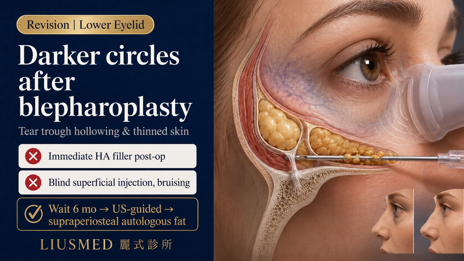
- The deepened shadow is not pure pigmentation — it stems from three anatomical changes: thinned translucent skin, tear-trough (hollow groove under lower eyelid) step-off, and post-surgical adhesion.
- Autologous fat grafting (Fat Grafting / Lipofilling — autologous fat transfer) can simultaneously restore volume, thicken the dermis, and improve skin quality, supported by international plastic surgery literature as a viable revision option.
- Periocular revision depends heavily on high-resolution ultrasound guidance — used not only for diagnosis, but as an intraoperative navigation tool to avoid vessels and locate adhesion planes.
- Not every case of worsening dark circles needs immediate intervention. Wait at least 6 months post-op for tissue stabilization before deciding on revision, to avoid being misled by edema-stage findings.
Why Do Dark Circles Look Worse After Surgery That Was Supposed to Fix Them?
In clinic, I frequently meet anxious patients who underwent lower eyelid blepharoplasty hoping for brighter, smoother eyes — only to discover, after the swelling subsided, that the bags are gone but the dark circles look deeper than ever, with visible hollowing under the eyes.
If this is happening to you, please don't blame yourself or your aftercare. This is not rare clinically. Plastic and Reconstructive Surgery – Global Open has published a focused analysis of this complication and its repair strategy. The cause is rarely poor recovery — it's almost always rooted in changes to tissue planes and subcutaneous volume loss introduced during surgery itself.
Anatomical Causes of Worsening Dark Circles After Blepharoplasty
Traditional lower lid blepharoplasty — particularly approaches that emphasize aggressive fat resection — can quickly flatten orbital bags but often disrupt the precise periocular architecture. Anatomically, three factors typically converge to deepen dark circles:
1. Thinned Skin and Vascular Show-Through
After significant orbital fat removal, the supporting volume disappears, and lower-eyelid skin drapes directly over the orbicularis oculi muscle and dense subcutaneous vasculature. The reddish-purple of those vessels becomes visible through the skin — the classic "vascular dark circle." Patients with naturally thin periorbital skin show this most dramatically.
2. Structural Tear-Trough Shadow
Over-resection creates a step-off between the tear trough and the lid–cheek junction. Whenever light strikes from above, that step casts a sharp shadow under the eye. This is a purely physical light-and-shadow problem — no amount of brightening cream or pigment laser can resolve it.
3. Adhesion and Post-Inflammatory Hyperpigmentation
During wound healing, mild scar adhesion plus post-inflammatory hyperpigmentation (PIH — darkening of skin after inflammation or injury) can deepen local darkness. This component takes time to clarify — usually 6+ months — before it can be evaluated accurately.
Key Insight: Lower lid blepharoplasty should never be reduced to pure subtraction. Periocular rejuvenation depends on a balanced architecture of skin, muscle, and fat. Once over-resection collapses that support, hollowing and vascular show-through are the predictable consequences.
Why Autologous Fat Grafting? How It Differs from HA (Hyaluronic Acid) Filler
When facing post-blepharoplasty volume loss, many patients first consider hyaluronic acid (HA — sugar molecule naturally in skin, holds water**) filler**. But the under-eye is an extremely thin, sensitive zone. The wrong material or the wrong injection plane easily produces the Tyndall effect (a bluish hue) or HA encapsulation and under-eye filler granuloma.
By contrast, autologous fat grafting offers material-level advantages in this exact context:
| Comparison | Autologous Fat Grafting | Hyaluronic Acid (HA) Fillers |
|---|---|---|
| Source | Patient's own adipocytes — no rejection or foreign-body reaction | Synthetic gel; may trigger delayed inflammation or capsule formation |
| Regenerative effect | Rich in adipose stem cells (ADSC — Adipose-Derived Stem Cells) and growth factors — improves skin quality, thickens the dermal barrier, reduces vascular show-through | Pure volumetric filler with no cellular regenerative effect |
| Visual naturalness | Once integrated, blends with surrounding tissue; no Tyndall blue | If placed too superficially, easily creates unnatural bumps or bluish discoloration |
| Long-term stability | Once survived, mostly long-term stable | Requires repeat sessions; cumulative injections can lead to residue and calcified nodules |
That said, autologous fat is not a universal fix-all. Improper technique, wrong injection plane, or excess volume in a single session can themselves trigger calcification, lumps, or surface irregularities (see autologous fat complication revision). The material's advantage only materializes when paired with precise injection technique.
A Surgeon's View: "If You Can't See It, You Can't Treat It Safely" — Ultrasound Guidance
Although autologous fat grafting is an excellent option for post-blepharoplasty dark circles, the injection plane decides safety and success. The periocular region is densely packed with microvasculature and nerves. "Blind" injection cannot accurately read the adhesion patterns left by prior surgery — and worse, accidental intravascular fat embolization can cause severe complications.
At our clinic we hold to one principle: "Only what you can see can be treated safely." Throughout the procedure I use high-resolution ultrasound. Importantly, ultrasound here is not just a diagnostic tool — it serves as a real-time intraoperative navigation tool for safety and precision.
Live ultrasound visualization lets me identify:
- The thickness and position of any residual fat pads
- Adhesion areas left by prior surgery
- Critical vessels (e.g., angular vessel, infraorbital vessel)
- Real-time needle-tip depth across tissue layers
This allows precise placement of microfat or nanofat into the correct fascial planes. The result: dramatically lower risk and better blood supply for grafted fat — higher survival rate and a smoother, more natural contour.
Key Insight: The best periocular revision is not driven by automated devices but by the surgeon's deep anatomical understanding paired with delicate hand technique. High-resolution ultrasound is our navigation radar — every advance is grounded and safe.
What Patients Need to Know About the Procedure
If you're considering full-face autologous fat grafting to repair post-blepharoplasty hollowing, here are the clinically critical points:
Timing: Wait at Least 6 Months Post-Op
Intervening before tissues have stabilized risks unpredictable interactions with residual edema or scar formation. We typically recommend at least 6 months from the prior blepharoplasty before evaluating revision — once anatomy and pigmentation are stable.
Pre-Operative Precision Assessment
- High-resolution ultrasound to map lower-lid thickness and blood flow
- Differentiating dark-circle etiology: vascular show-through vs. structural hollowing vs. pigmentation (each requires a different strategy)
- Mapping the extent of prior surgical adhesion
Fat Harvest and Purification
Small-volume fat is typically harvested from the abdomen or thigh, then purified to isolate the most refined growth-factor-rich microfat or even nanofat. The required refinement for the periocular zone exceeds that of standard facial filling.
Ultrasound-Guided Placement
Under local anesthesia, ultrasound guidance avoids danger zones; a blunt cannula deposits microfat in small aliquots, multiple passes, multi-layer distribution into hollows and the deep dermis.
Recovery
Mild swelling for the first three days, generally resolving in one to two weeks. During this period:
- Avoid rubbing the eye area
- Cold compress for the first 48 hours to reduce edema
- Avoid prone sleeping and saunas
- Give grafted fat the stability it needs to integrate
Restoring Bright Eyes Begins with Precise Assessment
Worsening dark circles and hollowing after blepharoplasty are deeply frustrating — but they are not irreversible. Anatomically grounded autologous fat grafting, paired with ultrasound-guided safety navigation, can restore lost volume and leverage fat's regenerative properties to thicken thinned periocular skin.
Every pair of eyes is unique. Repairing prior surgical adhesion and volume loss demands a highly individualized strategy. If post-blepharoplasty changes are causing you appearance-related distress, please don't rush into blind injections.
Come into clinic and let us perform a thorough ultrasound-based periocular evaluation, and design the safest, most effective revision plan together.
👉 Book a consultation: let Dr. Ta-Ju Liu design your personalized periocular revision plan
Medical References
- Autologous Fat Grafting for Dark Circle Exacerbation Following Lower Eyelid Blepharoplasty. Plastic and Reconstructive Surgery – Global Open. 2024.
- Coleman SR (Systematic Review). Structural Fat Grafting: Beyond the Lipocyte. Plast Reconstr Surg. 2006.
- The Current State of Fat Grafting: A Review of Harvesting, Processing, and Injection Techniques. Plast Reconstr Surg. 2015. PMID (PubMed Identifier): 26086386.
- Improving the Retention of Low-Volume Autologous Fat Grafting: A Comparative Analysis. PMC11249923. 2024.
- Surgical Anatomy of the Lower Eyelid Relevant to Lower Blepharoplasty. Facial Plast Surg Clin North Am. 2016.
- Tyndall Effect After Hyaluronic Acid Injection in the Tear Trough: Mechanism and Management. Aesthet Surg J. 2020.
Editorial Review: Reviewed by Dr. Ta-Ju Liu. Last reviewed: 2026-05-06. This article is educational and does not constitute individual medical advice; treatment decisions must be based on individualized anatomical evaluation.
Related Services

Specialties
Credentials
- Kaohsiung Medical University, School of Medicine
- Attending Physician, Dermatology, Kaohsiung Chang Gung Memorial Hospital
- Attending Physician, Aesthetic Center, Kaohsiung Chang Gung Memorial Hospital
- Visiting Physician, Dermatology, Xiamen Chang Gung Hospital
- Visiting Physician, Aesthetic Center, Xiamen Chang Gung Hospital
"For every surgery, I strive to achieve a good outcome through a small incision and refined technique. Minimally invasive surgery is not just a technique — it's a commitment of respect to every patient."
Recovery from filler complications needs peer support too
Want to learn more?
Schedule a consultation for professional evaluation and advice