IPL Reduces Redness but Thins Your Skin? The MMP-9 Connection You Need to Know
Intense pulsed light has been the workhorse of rosacea management for over two decades. Walk into almost any dermatology clinic with rosacea complaints, and IPL (Intense Pulsed Light) will likely be among the first treatments recommended. It works — redness decreases, visible vessels fade, and patients leave satisfied. For many, it becomes a regular maintenance procedure, performed every few months year after year.
But some patients who have been on this IPL treadmill for years begin to notice something troubling: their skin feels thinner, more fragile, and paradoxically more reactive than when they started. Products that used to absorb comfortably now sting. The skin bruises more easily. Fine vessels that were never visible before now show through what seems like increasingly translucent tissue. They wonder if they are imagining things, or if something has genuinely changed.
They are not imagining it. And the explanation involves a family of enzymes called matrix metalloproteinases — specifically MMP (Matrix Metalloproteinase)-9.
Table of Contents
- How IPL Works on Rosacea: The Intended Mechanism
- Matrix Metalloproteinases: The Enzymes That Remodel Your Skin
- The MMP-9 Upregulation Problem with Repeated IPL
- Clinical Evidence: What Happens to Skin Thickness Over Time
- IPL Treatment Frequency and Cumulative Risk: A Framework
- Protecting Your Dermal Matrix While Managing Rosacea Redness
How IPL Works on Rosacea: The Intended Mechanism {#how-ipl-works}
Unlike lasers that emit a single wavelength, IPL devices produce broad-spectrum light — typically spanning 500 to 1200 nm — filtered through cutoff filters to select specific wavelength bands. For rosacea, filters in the 515–600 nm range are commonly used to target oxyhemoglobin in dilated blood vessels.
The broadband nature of IPL means that multiple chromophores absorb energy simultaneously. While oxyhemoglobin is the primary target, melanin, water, and various tissue proteins also absorb portions of the spectrum. This makes IPL less selective than dedicated vascular lasers like pulsed dye laser but more versatile — it can address redness, pigmentation, and mild textural concerns in a single session.
For rosacea patients, the treatment workflow is straightforward: the IPL handpiece delivers pulses of filtered light across the affected areas. Hemoglobin in dilated vessels absorbs the light, the resulting heat damages the vessel endothelium, and the body's immune system clears the damaged vessels over the following days to weeks.
The treatment is effective. Studies consistently show 40–60% reduction in background erythema and significant reduction in visible telangiectasia after a series of three to five sessions. Patient satisfaction is high, at least initially.
The question that has received insufficient attention is: what is happening to the tissue between and around those vessels with each successive treatment?
Matrix Metalloproteinases: The Enzymes That Remodel Your Skin {#matrix-metalloproteinases}
Matrix metalloproteinases (MMPs) are a family of zinc-dependent enzymes responsible for breaking down components of the extracellular matrix (ECM) — the structural scaffold that gives skin its strength, elasticity, and thickness. Under normal circumstances, MMP activity is tightly regulated by their natural inhibitors, the tissue inhibitors of metalloproteinases (TIMPs).
The balance between MMPs and TIMPs determines whether the ECM is being maintained (balance), built up (TIMP dominance), or broken down (MMP dominance). In healthy skin, this balance favors maintenance, with controlled MMP activity during wound healing and remodeling.
Key MMPs relevant to skin structure include:
MMP-1 (collagenase-1): Cleaves fibrillar collagens (types I and III), the primary structural proteins of the dermis.
MMP-2 (gelatinase A): Degrades denatured collagen (gelatin) and basement membrane collagen (type IV).
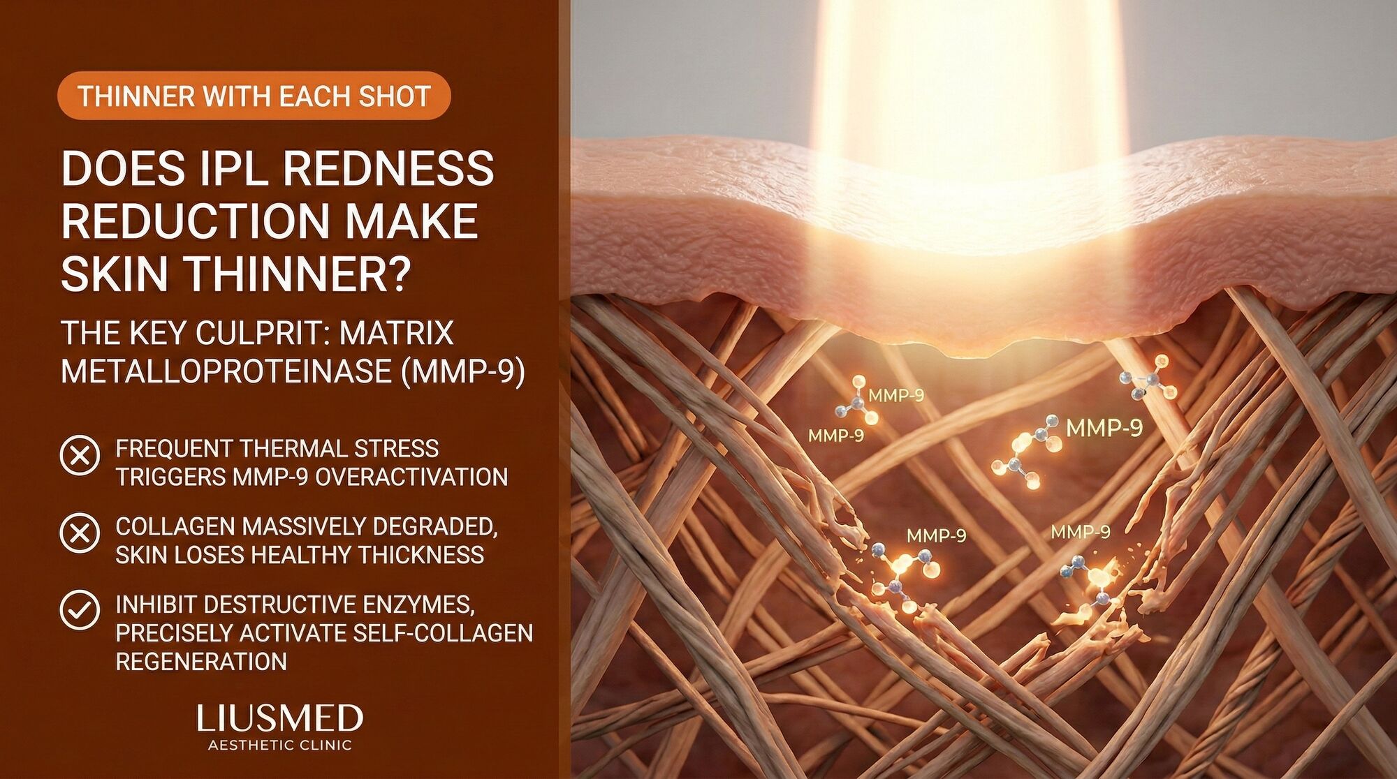
MMP-9 (gelatinase B): Degrades type IV collagen (basement membrane), type V collagen, elastin, and gelatin. Also releases matrix-bound growth factors including VEGF (Vascular Endothelial Growth Factor — new blood vessel signal).
MMP-3 (stromelysin-1): Broad substrate specificity; activates other MMPs and degrades proteoglycans, fibronectin, and laminin.
In rosacea, MMP levels — particularly MMP-2 and MMP-9 — are already elevated compared to healthy skin. This baseline elevation is part of the disease pathology and contributes to the dermal thinning and vascular instability that characterize the condition.
The critical question is: does IPL treatment increase or decrease MMP activity in rosacea skin?
The MMP-9 Upregulation Problem with Repeated IPL {#mmp9-upregulation}
The interaction between broad-spectrum light energy and MMP expression is well-documented in photobiology research, although much of this research focuses on ultraviolet radiation rather than visible/near-infrared IPL wavelengths. The key finding, consistently replicated across study designs, is that light-induced tissue heating and the resulting inflammatory response upregulate MMP production — particularly MMP-9.
The mechanism proceeds through several pathways:
Direct thermal activation. Heat stress in dermal fibroblasts activates the AP-1 transcription factor pathway, which directly upregulates MMP-1, MMP-3, and MMP-9 gene expression. The temperatures reached in perivascular tissue during IPL treatment (45–65 degrees Celsius) are well within the range that activates this pathway.
Inflammatory cytokine cascade. The tissue damage from IPL triggers release of IL-1beta and TNF-alpha from keratinocytes and immune cells. Both cytokines are potent inducers of MMP-9 expression in fibroblasts and inflammatory cells. In rosacea skin, where baseline cytokine levels are already elevated, this additional stimulus pushes MMP-9 expression to higher levels than would occur in healthy tissue.
Neutrophil-derived MMP-9. Neutrophils are among the first immune cells recruited to IPL-treated tissue. They carry pre-formed MMP-9 in their granules and release it upon activation. Since rosacea skin recruits neutrophils more aggressively than healthy skin, IPL treatment in rosacea generates a larger neutrophil-derived MMP-9 spike.
Mast cell-mediated MMP release. Activated mast cells release not only histamine but also MMP-9 and other proteases. Given the heightened mast cell reactivity in rosacea, IPL-triggered degranulation contributes significantly to post-treatment MMP-9 levels.
The cumulative effect of these pathways is a sustained elevation of MMP-9 activity in IPL-treated rosacea skin that extends well beyond the immediate post-treatment period. While a single session produces a transient spike that resolves within one to two weeks, repeated sessions at monthly intervals can maintain chronically elevated MMP-9 levels — each new spike occurring before the previous one has fully resolved.
Clinical Evidence: What Happens to Skin Thickness Over Time {#clinical-evidence}
The relationship between repeated IPL and dermal thinning in rosacea has been less studied than it deserves, partly because the effect is gradual and difficult to measure without specialized imaging. However, several lines of evidence support the connection:
Ultrasound measurement studies in patients receiving serial IPL treatments have documented measurable decreases in dermal thickness over 12 to 24 months of treatment, particularly in patients receiving more than four sessions per year.
Histological observations from biopsy samples of rosacea skin with extensive IPL treatment history show characteristic changes: reduced collagen bundle density, fragmented elastic fibers, thinned basement membranes around vessels, and expanded extracellular space (consistent with matrix degradation without adequate replacement).
Clinical correlation between treatment frequency and perceived skin fragility is consistently reported. Patients who have undergone more than fifteen to twenty IPL sessions over several years report significantly higher rates of skin thinning symptoms — increased bruising tendency, visible underlying vessels that were not previously apparent, stinging with previously tolerated products, and a translucent quality to the skin.
The pattern is consistent with what MMP-9 upregulation would predict: progressive degradation of the dermal matrix, particularly the basement membrane (type IV collagen) and elastic fiber network, leading to thinner, less resilient, more vascular-transparent skin.
| Indicator | Before IPL Treatment | After 5-10 Sessions | After 15+ Sessions |
|---|---|---|---|
| Dermal thickness | Baseline (may already be reduced in rosacea) | Minimal change or slight reduction | Measurable reduction in many patients |
| Bruising tendency | Normal or slightly increased | Similar to baseline | Often noticeably increased |
| Product sensitivity | Moderate (typical rosacea) | Similar to baseline | Frequently increased |
| Vessel visibility | Dilated superficial vessels | Reduced (treatment working) | New, finer vessels visible through thinner skin |
| MMP-9 levels | Elevated (rosacea baseline) | Transiently spiking with each session | Chronically elevated with incomplete recovery |
| Elastic fiber integrity | Partially degraded | Progressive fragmentation | Significant loss of recoil |
| Basement membrane | Mildly thinned | Progressive thinning | Compromised structural support |
IPL Treatment Frequency and Cumulative Risk: A Framework {#treatment-frequency-risk}
Not all IPL usage carries the same risk of matrix degradation. The variables that determine cumulative impact include treatment frequency, fluence (energy level), pulse duration, number of passes per session, filter selection, and the baseline inflammatory state of the skin.
A risk-stratified framework:
Lower risk profile: Quarterly IPL sessions (4 per year or fewer), conservative fluences, single pass per area, on skin that has been pre-treated with anti-inflammatory agents to reduce baseline MMP-9 and cytokine levels. This approach limits cumulative MMP-9 exposure while still providing meaningful vascular treatment.
Moderate risk profile: Monthly sessions during an initial treatment series (4–6 sessions), followed by quarterly maintenance. Moderate fluences with occasional double-pass on resistant vessels. This is the most common clinical protocol and carries meaningful cumulative risk over multiple years of treatment.
Higher risk profile: Monthly or more frequent sessions continued indefinitely, aggressive fluences, multiple passes per session, on actively inflamed skin without anti-inflammatory pre-treatment. This protocol maximizes MMP-9 upregulation at each session while providing insufficient recovery time between treatments.
The distinction between these profiles highlights an important principle: IPL itself is not the enemy. The problem is the application pattern — specifically, frequency and intensity that exceeds the tissue's capacity to recover and rebalance its MMP/TIMP ratio between sessions.
Protecting Your Dermal Matrix While Managing Rosacea Redness {#protecting-dermal-matrix}
For rosacea patients who need vascular treatment, the goal should be achieving redness reduction while minimizing cumulative matrix degradation. Several strategies support this balance:
Pre-treatment anti-inflammatory preparation. Reducing baseline inflammation before IPL decreases the inflammatory amplification that drives excessive MMP-9 production. This may involve topical anti-inflammatory agents, oral anti-inflammatory medications, or targeted interventions like Rosacea Injection Treatment to calm the perivascular microenvironment before any light-based treatment is applied.
Extended intervals between sessions. Allowing a minimum of eight to twelve weeks between IPL sessions — rather than the commonly prescribed four weeks — gives MMP-9 levels time to normalize and the tissue time to rebalance its matrix turnover. The cosmetic result may take longer to achieve, but the structural cost is substantially lower.
Post-treatment MMP modulation. Topical agents with demonstrated MMP-inhibitory activity — including certain retinoids (which paradoxically increase MMP-1 but decrease MMP-9 at appropriate concentrations), niacinamide, and specific botanical extracts with MMP-inhibitory properties — can help counteract the post-IPL MMP spike.
Matrix support supplementation. Supporting the raw materials for matrix repair — vitamin C for collagen synthesis, specific amino acids, and compounds that support glycosaminoglycan production — provides the building blocks needed to replace matrix components degraded by elevated MMPs.
Regular monitoring. Clinical assessment of skin thickness, sensitivity, and bruising tendency should be part of every IPL follow-up. If these parameters are trending negatively, treatment frequency and intensity should be reduced regardless of the cosmetic result.
Treatment endpoint definition. Perhaps most importantly, rosacea patients and their practitioners should define a stopping point or maintenance protocol before beginning IPL treatment. Open-ended "keep treating until it is perfect" approaches are where the highest cumulative risk accumulates. Defining "good enough" and transitioning to non-IPL maintenance strategies protects the dermal matrix from indefinite degradation.
The MMP-9 connection is not a reason to categorically refuse IPL treatment. For many rosacea patients, the functional and psychological benefits of reduced redness justify the treatment, provided it is delivered thoughtfully. But understanding the mechanism of cumulative matrix degradation allows patients and practitioners to make informed decisions about treatment frequency, intensity, duration, and the importance of matrix-protective adjunctive care.
Your skin is not just a surface to be treated — it is a living structural matrix that must support you for decades. Every treatment decision should weigh immediate cosmetic benefit against long-term structural integrity.
Frequently Asked Questions
Q1: Can I test my MMP-9 levels to know if IPL is damaging my skin?
While MMP-9 can be measured through skin biopsy with immunohistochemistry or through analysis of wound fluid, these tests are not part of routine clinical practice for aesthetic patients. A more practical approach is clinical monitoring: tracking skin thickness (which can be assessed with high-frequency ultrasound), sensitivity, bruising tendency, and vessel visibility between sessions. Trending negatively on these clinical measures is a reasonable proxy for excessive MMP activity.
Q2: I have been getting IPL monthly for two years and my skin feels thinner. Is the damage permanent?
Matrix degradation from chronic MMP elevation is partially reversible with appropriate intervention. The collagen component can be restored through stimulating fibroblast activity, although this takes months to years of consistent support. Elastic fiber loss is more difficult to reverse — adult skin has limited capacity for elastin synthesis. Stopping the excessive MMP stimulus (reducing IPL frequency), supporting matrix repair, and allowing recovery time will produce improvement, but the timeline is measured in six to eighteen months for significant structural recovery.
Q3: Are there IPL devices that produce less MMP-9 upregulation than others?
Devices with shorter pulse durations, better cooling systems, and more precise filter options that minimize non-target wavelength delivery may produce less collateral tissue heating and therefore less MMP activation. However, the fundamental mechanism — tissue heating leading to inflammatory cytokine release leading to MMP upregulation — applies to all IPL devices. Device selection matters less than treatment parameters and frequency.
Q4: Does MMP-9 from IPL also affect the blood vessels themselves, making them less stable?
Yes. MMP-9 degrades type IV collagen in the vascular basement membrane, directly weakening vessel walls. This means that while IPL destroys existing dilated vessels, the MMP-9 spike it triggers simultaneously weakens the structural support of remaining and newly forming vessels. This creates a paradox: treatment that closes some vessels while destabilizing others, potentially contributing to the formation of new telangiectasia in adjacent areas.
Q5: My practitioner says newer IPL technology does not cause these problems. Is that accurate?
Newer IPL platforms have improved in several ways: better cooling, more refined filter options, and improved pulse-shaping technology. These advances reduce collateral thermal damage and improve target selectivity. However, they do not eliminate the fundamental inflammatory response to broad-spectrum light in chronically inflamed tissue. Reduced collateral damage means reduced MMP activation — which is beneficial — but not zero MMP activation. The cumulative principle still applies, particularly over many sessions.
Q6: Should I take oral supplements to inhibit MMP-9 while getting IPL treatments?
Certain oral supplements have demonstrated MMP-inhibitory properties in laboratory studies, including green tea catechins (EGCG), omega-3 fatty acids, and curcumin. However, systemic MMP inhibition carries its own risks — MMPs are essential for wound healing, immune function, and tissue remodeling throughout the body. Targeted, local approaches to MMP modulation in treated skin are generally preferable to systemic supplementation. Discuss any supplement regimen with your treating physician.
About the Author
Dr. Ta-Ju Liu is the founder of Liusmed Clinic, specializing in regenerative medicine and minimal incision surgery. Dr. Liu's approach to rosacea management integrates structural dermal assessment with vascular treatment planning, ensuring that interventions designed to reduce redness do not inadvertently compromise the dermal matrix. Liusmed Clinic offers comprehensive rosacea protocols that balance cosmetic outcomes with long-term skin structural integrity, serving patients from Taiwan and internationally.
Disclaimer
This article is provided for educational and informational purposes only and does not constitute medical advice, diagnosis, or treatment. IPL is an established medical technology with a strong safety record when used appropriately. This article discusses potential cumulative effects of repeated treatments specifically in the context of rosacea, a chronic inflammatory condition. Individual responses to treatment vary significantly. All treatment decisions should be made in consultation with a qualified healthcare provider.

Specialties
Credentials
- Kaohsiung Medical University, School of Medicine
- Attending Physician, Dermatology, Kaohsiung Chang Gung Memorial Hospital
- Attending Physician, Aesthetic Center, Kaohsiung Chang Gung Memorial Hospital
- Visiting Physician, Dermatology, Xiamen Chang Gung Hospital
- Visiting Physician, Aesthetic Center, Xiamen Chang Gung Hospital
"For every surgery, I strive to achieve a good outcome through a small incision and refined technique. Minimally invasive surgery is not just a technique — it's a commitment of respect to every patient."
Recovery after any procedure needs peer support too
Want to learn more?
Schedule a consultation for professional evaluation and advice