Visible Vessels & Hypersensitive Skin: a 'Laser Refugee'?
There is a term circulating among aesthetic dermatology patients in East Asia that captures a particular kind of suffering: "laser refugee." It describes someone who sought laser treatment for a cosmetic concern, usually melasma, and emerged with skin that is objectively worse than when they started. Not just the same pigmentation, but new problems layered on top: chronic sensitivity, flushing at the slightest trigger, visible spider veins across the cheeks, and a skin surface that feels thin, fragile, and permanently irritated.
These patients often move from clinic to clinic seeking help, hence the "refugee" label. Each new provider identifies the damage from previous treatments but may struggle to offer solutions that do not carry additional risk to already compromised skin.
If this describes your experience, understanding exactly what happened to your skin is the essential first step toward recovery.
Table of Contents
- What Is Laser Refugee Syndrome?
- The Three Layers of Damage: Barrier, Vasculature, and Melanocytes
- How Repeated Melasma Laser Treatments Create This Cascade
- Self-Assessment: Signs That Your Skin Has Been Over-Treated
- Conventional Laser Approach vs. Regenerative Repair: A Comparison
- The Road to Recovery: Rebuilding Damaged Skin
What Is Laser Refugee Syndrome?
Laser refugee syndrome is not a formal medical diagnosis but rather a descriptive term for the constellation of symptoms that develops after excessive laser treatment. The pattern is remarkably consistent across patients, regardless of the specific laser type used:
First comes increased baseline redness. The skin develops a persistent pinkish or reddish tone that was not present before treatment. This is not temporary post-procedural erythema; it persists for weeks and months, becoming the new normal.
Next comes heightened sensitivity. Products that were previously tolerated now cause stinging or burning. Temperature changes trigger flushing. Even gentle cleansing feels uncomfortable. The skin's tolerance threshold has been dramatically lowered.
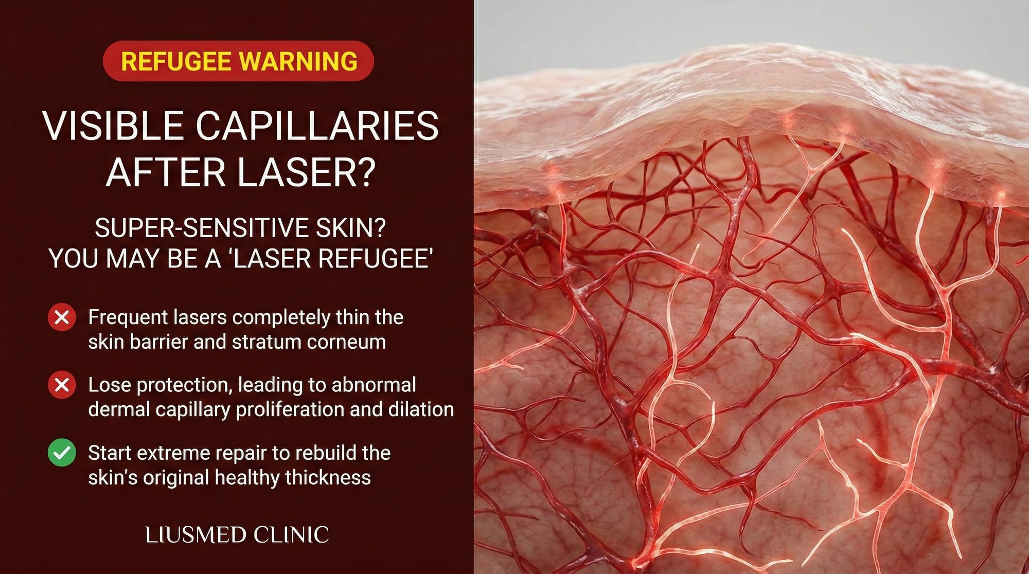
Then the vessels become visible. Fine red or purple blood vessels, medically termed telangiectasia, appear across the cheeks, nose, and sometimes the forehead. These represent dilated capillaries and venules that have lost their ability to properly constrict.
Throughout all of this, the original melasma often remains or has worsened. The patient now has two problems instead of one: the pigmentation they originally sought to treat, plus a damaged, sensitized skin barrier with vascular dysfunction.
The emotional impact is significant. Many laser refugees describe feeling betrayed by the aesthetic industry, anxious about their appearance, and uncertain whether their skin can ever return to its pre-treatment state. These feelings are valid and deserve acknowledgment alongside the physical treatment plan.
The Three Layers of Damage: Barrier, Vasculature, and Melanocytes
Understanding laser refugee syndrome requires recognizing that the damage occurs simultaneously across three interconnected systems.
The epidermal barrier. The stratum corneum, the outermost layer of skin, functions as a brick-and-mortar wall where corneocytes (dead skin cells) are the bricks and lipids (ceramides, cholesterol, and fatty acids) are the mortar. Repeated laser exposure disrupts this architecture in several ways. The thermal and mechanical energy damages corneocytes and depletes intercellular lipids. The accelerated cell turnover induced by laser does not allow adequate time for the barrier to fully reconstruct between sessions. Over multiple treatments, the barrier becomes progressively thinner and more permeable, explaining the increased sensitivity to products, temperature, and environmental irritants.
Transepidermal water loss (TEWL) measurements in patients with laser refugee syndrome consistently show values well above normal, confirming objective barrier dysfunction. This is not a subjective complaint; it is measurable structural damage.
The dermal vasculature. The dermis contains a complex network of blood vessels that serve multiple functions: nutrient delivery, temperature regulation, immune surveillance, and waste removal. These vessels are normally regulated by a balance of vasoconstrictive and vasodilatory signals.
Repeated laser energy delivery generates heat in the dermis that triggers vasodilation. When this occurs frequently, the vessel walls undergo structural changes. Smooth muscle cells in the vessel walls weaken, and the elastic fibers that allow vessels to constrict degrade. The result is permanently dilated vessels that are visible through the now-thinner epidermis.
Additionally, the chronic subclinical inflammation from repeated treatments upregulates vascular endothelial growth factor (VEGF), promoting the formation of new blood vessels (angiogenesis). This is particularly problematic in the context of melasma, as increased dermal vascularity is itself a driver of melanocyte activation.
The melanocyte population. Melanocytes can be damaged in two opposing directions. Some are destroyed, leading to small depigmented spots (guttate hypomelanosis or confetti-like depigmentation). Others are hyperactivated by the inflammatory milieu, producing more melanin than before. The result is a mottled, uneven appearance where areas of hyperpigmentation alternate with areas of hypopigmentation. This pattern is far more difficult to treat than the original uniform melasma.
How Repeated Melasma Laser Treatments Create This Cascade
The cascade typically unfolds over months or years of regular treatment. The early phase involves sessions one through five, where results appear positive and both patient and provider are encouraged. Any minor sensitivity or redness is dismissed as expected and temporary.
The middle phase, sessions five through fifteen, is where the damage accumulates below the threshold of clinical detection. TEWL increases. The dermal vascular density begins to change. Melanocyte behavior becomes increasingly unpredictable. Some patients notice that their skin flushes more easily or that their usual skincare products cause mild stinging.
The late phase, beyond fifteen sessions, is when the damage becomes clinically obvious. Persistent redness, visible vessels, chronic sensitivity, and the paradoxical worsening of pigmentation all converge into the laser refugee presentation.
Several factors accelerate this cascade. Shorter intervals between treatments do not allow adequate healing. Higher fluence settings increase the energy dose per session. Lack of proper skin assessment between sessions means early warning signs are missed. Failure to adjust or halt treatment in response to barrier compromise allows damage to compound.
The fundamental issue is that the treatment model prioritizes pigment clearance over skin health. Each session is evaluated based on whether the melasma looks lighter, with insufficient attention paid to what is happening to the rest of the skin's biology.
Self-Assessment: Signs That Your Skin Has Been Over-Treated
Consider whether you experience any of the following. The more items you identify with, the higher the likelihood that your skin has sustained cumulative laser damage:
Barrier compromise indicators: Your skin stings when applying products that previously caused no reaction. Your skin feels tight and dry within an hour of cleansing. You cannot tolerate any active skincare ingredients such as vitamin C, retinoids, or acids. Your skin peels or flakes easily with minimal friction.
Vascular dysfunction indicators: Your cheeks flush bright red in response to mild temperature changes, spicy food, alcohol, exercise, or emotional stress. You can see fine red or purple blood vessels on your cheeks, nose, or chin that were not there before. Your skin has a persistent pink or red undertone that does not resolve with rest. Hot showers or baths cause prolonged facial redness lasting more than thirty minutes.
Melanocyte disruption indicators: You have noticed small white spots (smaller than a pencil eraser) appearing on your cheeks or treatment areas. Your melasma appears more patchy or irregular than before treatment. Some areas seem darker while adjacent areas seem lighter, creating a mottled appearance. Your skin darkens rapidly and disproportionately with even minimal sun exposure.
Inflammatory indicators: Your skin frequently feels warm to the touch even without external triggers. You develop bumps, papules, or a rash-like texture in the treatment area. Your skin reacts to wind, cold, or air conditioning changes with burning sensations.
If you identify with items across multiple categories, your skin is likely in a compromised state that requires a recovery-focused approach rather than additional active treatments.
Conventional Laser Approach vs. Regenerative Repair: A Comparison
| Factor | Continued Laser Treatment | Regenerative Repair Strategy |
|---|---|---|
| Philosophy | Continue treating pigment despite barrier damage | Halt further damage; prioritize barrier and vascular recovery |
| Barrier Impact | Further disruption with each session | Active repair using barrier-restoring agents |
| Vascular Impact | Continued thermal stress on already-dilated vessels | Vascular normalization through anti-angiogenic modulation |
| Melanocyte Impact | Ongoing unpredictable stimulation and destruction | Gentle microenvironment modulation to reduce hyperactivation |
| Sensitivity Trajectory | Likely to worsen progressively | Gradual improvement as barrier function restores |
| Timeline | Indefinite ongoing treatment | Defined recovery phase (typically 6-12 months) |
| Risk Profile | High risk of further compounding damage | Low risk; focused on healing rather than active destruction |
| Long-term Prognosis | Continued deterioration without addressing root cause | Potential for meaningful recovery of skin function |
The critical distinction is between doing more and doing differently. For a laser refugee, more of the same treatment that caused the damage is contraindicated. The Melasma Injection Treatment approach offers an alternative that can address pigmentation without inflicting the thermal and mechanical trauma that worsens barrier and vascular dysfunction.
The Road to Recovery: Rebuilding Damaged Skin
Recovery from laser refugee syndrome is possible but requires patience, discipline, and a fundamentally different approach than what created the problem.
Phase one: Cessation and stabilization (months one through three). All laser and energy-based treatments must stop completely. The skincare routine should be stripped to bare essentials: a gentle, non-foaming cleanser, a barrier-repair moisturizer rich in ceramides and cholesterol, and a mineral-based broad-spectrum sunscreen. No active ingredients, no exfoliation, no treatment serums. The goal is to stop all assault on the barrier and allow the skin's intrinsic repair mechanisms to begin working.
Phase two: Active barrier repair (months three through six). Once the skin has stabilized and acute sensitivity has decreased, barrier-repair strategies can be introduced more aggressively. This may include prescription-grade barrier repair formulations, anti-redness ingredients like niacinamide and centella asiatica extracts, and targeted treatments such as Melasma Injection Treatment that deliver reparative agents directly to the dermis without further compromising the epidermis.
Phase three: Pigment management (months six through twelve). Only after the barrier has been substantially restored and vascular reactivity has decreased should pigment-specific treatments be considered. At this stage, the approach should be strictly non-thermal and non-ablative. Injectable therapies that deliver melanogenesis-modulating agents directly to the affected tissue can address the remaining pigmentation without risking a return to the inflammatory cycle.
Phase four: Maintenance and prevention (ongoing). Long-term maintenance includes consistent sun protection, gentle skincare, periodic assessment of barrier function and vascular status, and prompt intervention at the first sign of recurrence. The goal shifts from active treatment to preserving the gains achieved during recovery.
Throughout this process, realistic expectations are essential. Some vascular changes, particularly established telangiectasia, may not fully reverse without targeted vascular laser treatment, which can be cautiously considered once the skin has recovered. Some melanocyte damage, particularly depigmented spots, may be permanent. The overall trajectory should be toward significant improvement in skin quality, sensitivity, and appearance, even if perfection is not achievable.
Frequently Asked Questions
Q1: Can the visible blood vessels on my face be treated, or are they permanent?
Established telangiectasia (visible dilated blood vessels) can often be improved with vascular-specific laser treatments such as pulsed dye laser (PDL) or KTP laser at appropriate parameters. However, these treatments should only be considered after the skin barrier has been substantially restored, typically no sooner than six to twelve months after cessation of the treatments that caused the damage. Treating vessels in actively compromised skin risks further sensitization.
Q2: How long does it take for over-treated skin to recover?
The timeline depends on the severity of damage and the duration of over-treatment. Mild cases with primarily barrier dysfunction may show significant improvement within three to six months of proper care. Moderate cases involving barrier damage plus vascular changes typically require six to twelve months. Severe cases with all three layers of damage, involving barrier, vascular, and melanocyte disruption, may take twelve to eighteen months for meaningful recovery. Some changes may be partially irreversible.
Q3: My skin burns when I apply sunscreen. How am I supposed to protect it from the sun?
This is a common frustration for laser refugees. Start with a purely mineral (zinc oxide and/or titanium dioxide) sunscreen in a base formulated for sensitive skin, free of fragrance, essential oils, and chemical UV filters. If even this is not tolerated, use physical barriers such as a wide-brimmed hat, UV-blocking umbrella, and sun-protective clothing while your barrier repairs. As your skin heals, sunscreen tolerance typically improves over weeks to months.
Q4: Should I avoid all skincare products during recovery?
Not all products, but the routine should be radically simplified. Keep three products: a gentle cleanser, a barrier-repair moisturizer, and mineral sunscreen. Avoid all actives including vitamin C, retinoids, AHAs, BHAs, and benzoyl peroxide until your skin can tolerate its basic routine without any stinging, burning, or redness. Reintroduce active ingredients one at a time, with at least two weeks between additions, to identify any that your skin cannot yet tolerate.
Q5: Is my skin permanently damaged, or can it fully recover?
Most components of laser refugee syndrome are partially or fully reversible with appropriate care and time. Barrier function can be restored in virtually all cases. Vascular reactivity typically improves substantially, though some patients retain increased flushing tendency long-term. Visible telangiectasia may require targeted treatment but can often be significantly reduced. Melanocyte damage in the form of small depigmented spots may be permanent. The overall prognosis for meaningful recovery is good with the right approach and patience.
Q6: Can I get the melasma injection treatment while my skin is still sensitized?
The Melasma Injection Treatment approach is generally better tolerated than energy-based devices even in sensitized skin, because it does not generate thermal or mechanical trauma to the epidermal barrier. However, the timing of initiation depends on the severity of your skin compromise. In mild cases, injection therapy may begin during the active barrier repair phase. In severe cases, it may be advisable to wait until the barrier has substantially recovered. A thorough in-person assessment is necessary to determine the optimal timing for your specific situation.
About the Author
Dr. Ta-Ju Liu is the founder of Liusmed Clinic, a specialized center for regenerative medicine and minimal incision surgery in Taiwan. Dr. Liu has treated numerous patients presenting with cumulative skin damage from excessive laser therapy, developing structured recovery protocols that prioritize barrier restoration and vascular normalization before addressing residual pigmentation concerns. His dual expertise in dermatology and regenerative medicine informs a patient-centered approach to aesthetic complication repair.
Disclaimer
This article is provided for educational and informational purposes only and does not constitute medical advice. Individual skin conditions vary significantly, and treatment outcomes depend on numerous patient-specific factors. Always consult a qualified dermatologist or medical professional before making decisions about melasma treatment or skin recovery. The information presented here reflects current medical understanding as of the publication date and may be updated as new research becomes available.

Specialties
Credentials
- Kaohsiung Medical University, School of Medicine
- Attending Physician, Dermatology, Kaohsiung Chang Gung Memorial Hospital
- Attending Physician, Aesthetic Center, Kaohsiung Chang Gung Memorial Hospital
- Visiting Physician, Dermatology, Xiamen Chang Gung Hospital
- Visiting Physician, Aesthetic Center, Xiamen Chang Gung Hospital
"For every surgery, I strive to achieve a good outcome through a small incision and refined technique. Minimally invasive surgery is not just a technique — it's a commitment of respect to every patient."
Recovery after any procedure needs peer support too
Want to learn more?
Schedule a consultation for professional evaluation and advice