Six Months of Lightening Creams with No Results? Your Melasma May Have "Dropped" to the Dermis
You have been disciplined. Every morning: cleanser, vitamin C serum, hydroquinone, sunscreen. Every night: retinoid, azelaic acid, moisturizer. Six months in, you tilt your face toward the bathroom mirror, searching for improvement. The patches look exactly the same — or perhaps even slightly darker. Before concluding that your skin is uniquely resistant to treatment or that the products are ineffective, consider a possibility that most over-the-counter skincare marketing never mentions: the melanin causing your melasma may no longer be in a location that topical products can reach. It may have dropped into the dermis.
Table of Contents
- Epidermal vs. Dermal Melanin: The Depth Problem
- How Melanin "Drops" into the Dermis
- Why Topical Agents Cannot Reach Dermal Pigment
- Identifying Dermal Melasma: Clinical Clues and Diagnostic Tools
- The Paradox of Aggressive Topical Treatment
- What Actually Works for Dermal Melasma
Epidermal vs. Dermal Melanin: The Depth Problem
To understand why your lightening cream might be failing, you need to understand skin architecture. The skin consists of two primary layers: the epidermis (the outer layer, approximately 0.1 mm thick on the face) and the dermis (the deeper structural layer, 1 to 4 mm thick). Melanocytes — the cells that produce melanin — reside at the very bottom of the epidermis, at the junction between these two layers known as the dermal-epidermal junction (DEJ).
Under normal circumstances, melanin produced by melanocytes is packaged into melanosomes and transferred upward to keratinocytes in the epidermis. As these keratinocytes migrate toward the skin surface through the natural turnover process, the melanin is eventually shed. This is why a suntan fades over weeks — the pigmented keratinocytes are gradually replaced by new, less pigmented cells from below.
Epidermal melasma follows this model. The excess melanin sits within the epidermal keratinocytes, and treatments that suppress melanin production (hydroquinone, arbutin) or accelerate epidermal turnover (retinoids, chemical peels) can effectively reduce the visible pigmentation. The treatment is working within the same compartment where the pigment resides.
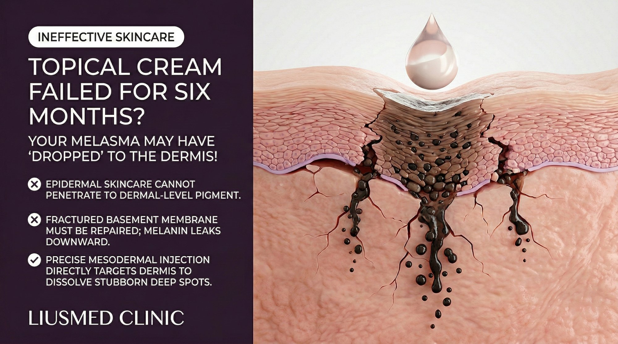
Dermal melasma represents a fundamentally different scenario. Here, melanin has crossed the DEJ and been deposited in the dermis — a compartment that does not undergo the same turnover process as the epidermis. Dermal melanin is engulfed by dermal macrophages (melanophages), which can retain the pigment for months to years. This melanin is no longer accessible to the natural shedding process, and it sits below the reach of most topical formulations.
The colour difference between epidermal and dermal melanin is clinically significant. Epidermal melanin appears brown — the melanin is close to the surface and absorbs light directly. Dermal melanin appears blue-grey or slate-grey because the Tyndall effect scatters shorter wavelengths of light as they pass through the overlying epidermis. If your melasma patches have a grey-blue tint rather than a warm brown tone, dermal involvement is likely.
How Melanin "Drops" into the Dermis
Melanin dropout — also called pigmentary incontinence — occurs when the dermal-epidermal junction is damaged or disrupted, allowing melanin granules and even entire melanocytes to fall into the dermis. Several mechanisms can cause this.
Chronic inflammation is the most common driver. When the basal layer of the epidermis is chronically inflamed, the structural proteins that anchor it to the dermis — including type IV collagen and various laminins — become degraded by matrix metalloproteinases (MMPs) released by inflammatory cells. As the junction weakens, melanin-laden cells and free melanin granules leak through the compromised barrier into the dermis below.
UV radiation damage contributes to DEJ disruption through both direct structural damage and indirect inflammatory pathways. Chronic UV exposure degrades the basement membrane zone and generates reactive oxygen species that further compromise junction integrity. This is one reason why melasma tends to progress from an epidermal to a mixed or dermal phenotype over time in patients with ongoing sun exposure.
Aggressive topical treatments can paradoxically accelerate melanin dropout. High-concentration chemical peels, overly potent retinoid formulations, and prolonged hydroquinone use can cause chronic low-grade inflammation at the DEJ. The very agents intended to lighten the skin may be pushing melanin deeper by damaging the barrier that normally keeps it contained in the epidermis.
Hormonal factors also play a role. Elevated estrogen has been shown to influence basement membrane protein expression, potentially weakening the DEJ in hormonally susceptible individuals. This may explain why pregnancy-associated melasma frequently develops a dermal component — the combination of hormonal DEJ weakening and increased melanocyte activity creates ideal conditions for pigmentary incontinence.
Once melanin enters the dermis, it is engulfed by macrophages that take up residence in the papillary and upper reticular dermis. These melanophages are remarkably persistent — they can retain melanin pigment for years. Even after the original melanocyte hyperactivity is controlled, the melanin already deposited in the dermis remains visible and is extremely difficult to clear.
Why Topical Agents Cannot Reach Dermal Pigment
The skin's barrier function — the very property that protects you from environmental hazards — is also the reason topical agents fail against dermal pigment. The stratum corneum, the outermost layer of the epidermis, is designed to prevent molecules from penetrating into the body. Only molecules with very specific properties can traverse it: low molecular weight (typically under 500 Daltons), moderate lipophilicity, and lack of electrical charge.
Even molecules that do penetrate the stratum corneum face a secondary challenge: concentration gradient. As a topical agent diffuses deeper, its concentration drops exponentially. By the time any active ingredient reaches the dermal-epidermal junction — let alone the dermis where melanophages reside — the concentration is a fraction of what was applied to the surface. For most depigmenting agents, this residual concentration is below the therapeutic threshold needed to affect dermal melanin.
Consider the specific agents commonly used for melasma:
| Topical Agent | Molecular Weight | Primary Site of Action | Effective Penetration Depth | Can Reach Dermis? |
|---|---|---|---|---|
| Hydroquinone | 110 Da | Melanocytes at DEJ | Upper epidermis to DEJ | Marginal — insufficient concentration |
| Tretinoin | 300 Da | Keratinocytes, DEJ | Epidermis | No meaningful dermal effect |
| Ascorbic acid (Vit C) | 176 Da | Epidermis | Upper epidermis | No — degrades before reaching dermis |
| Azelaic acid | 188 Da | Melanocytes | Epidermis to upper DEJ | Minimal |
| Kojic acid | 142 Da | Tyrosinase in epidermis | Epidermis | No |
| Tranexamic acid (topical) | 157 Da | Plasmin in epidermis | Epidermis to DEJ | Poor — oral form is far more effective |
| Niacinamide | 122 Da | Melanosome transfer | Epidermis | No |
The table reveals a consistent pattern: every commonly used topical depigmenting agent exerts its primary effect within the epidermis. None of them can meaningfully reduce melanin that has already been deposited in the dermis. This is not a product quality issue or a matter of using the wrong brand — it is a fundamental pharmacokinetic limitation.
Identifying Dermal Melasma: Clinical Clues and Diagnostic Tools
Determining whether your melasma has a dermal component is the single most important diagnostic step for predicting treatment response. Several methods can help.
Wood lamp examination is the classic first-line tool. Under 365 nm UV light, epidermal melanin absorbs the radiation and appears darker — the lesion boundary becomes more prominent against surrounding skin. Dermal melanin does not enhance under Wood lamp because the overlying epidermis scatters the UV light before it reaches the deeper pigment. If your melasma patches show strong enhancement under Wood lamp, the pigment is primarily epidermal and should respond to topical therapy. If they show minimal or no enhancement, significant dermal involvement is present.
Clinical colour assessment provides a useful approximation. Brown melasma with well-defined borders suggests epidermal pigment. Grey-blue or slate-coloured melasma with diffuse, poorly defined borders suggests dermal pigment. Most cases are mixed, with varying proportions of epidermal and dermal melanin across different areas of the same patch.
Dermatoscopic examination can reveal the depth and pattern of pigmentation. Epidermal melanin appears as a regular brown network. Dermal melanin creates a structureless blue-grey haze. The presence of reticular vascular patterns beneath the pigmentation suggests an active vascular-inflammatory component contributing to ongoing pigment dropout.
Duration of treatment resistance is itself a diagnostic clue. If you have used evidence-based topical depigmenting agents consistently for six months or longer without meaningful improvement, dermal pigment involvement should be assumed until proven otherwise. Epidermal melasma typically shows at least partial response within 8 to 12 weeks of appropriate topical therapy.
Reflectance confocal microscopy provides the most detailed non-invasive assessment, allowing direct visualization of melanin-laden cells at different skin depths. Where available, this technology can precisely quantify the epidermal-to-dermal melanin ratio and guide treatment selection accordingly.
The Paradox of Aggressive Topical Treatment
When topical treatments fail, the instinctive response is to escalate — higher concentrations, more frequent application, longer treatment duration, or addition of multiple active agents simultaneously. This escalation can be counterproductive for two reasons.
First, irritant dermatitis from overuse of active ingredients creates chronic inflammation at the DEJ. This inflammation degrades the basement membrane and promotes further pigmentary incontinence — pushing more melanin from the epidermis into the dermis. The patient perceives the worsening as treatment failure and escalates further, creating a vicious cycle. High-concentration hydroquinone (above 4 percent) used for extended periods is a particular offender, with the potential to cause not only increased pigment dropout but also ochronosis — a paradoxical permanent blue-black discoloration.
Second, aggressive exfoliation (through strong chemical peels or intensive retinoid protocols) strips the epidermal melanin efficiently but does nothing about the dermal component. The result is a confusing clinical picture: the skin appears lighter immediately after treatment, giving a false sense of progress, but the improvement reverses within days to weeks as the remaining dermal melanin becomes more visually prominent once the overlying epidermal pigment is removed. This phenomenon — where the skin looks temporarily better then relapses — is almost pathognomonic for mixed or dermal melasma.
The rational approach is to stop escalating topical therapy once dermal involvement is identified and to pivot to treatment modalities that can reach the dermis.
What Actually Works for Dermal Melasma
Managing dermal melasma requires accepting two realities: the treatment timeline is longer than for epidermal melasma, and the therapeutic approach must shift from topical suppression to systemic and interventional strategies.
Oral tranexamic acid is currently the most evidence-supported systemic treatment for dermal melasma. Unlike topical tranexamic acid, the oral form achieves therapeutic concentrations in the dermis through systemic circulation. It reduces plasmin activity, which in turn decreases prostaglandin production, VEGF (Vascular Endothelial Growth Factor — new blood vessel signal) expression, and melanocyte-stimulating inflammatory signals. Multiple randomized controlled trials have demonstrated significant improvement in melasma severity indices with oral tranexamic acid at doses of 250 mg twice daily.
Rigorous photoprotection with tinted mineral sunscreen remains the non-negotiable foundation. Preventing further UV-induced inflammation and DEJ damage stops the progression of melanin dropout even though it cannot reverse existing dermal deposits.
Targeted injection therapies represent the most direct approach to dermal melasma. Melasma Injection Treatment delivers active agents directly to the dermal layer where melanophages reside and where the vascular-inflammatory signals originate. By bypassing the epidermal barrier entirely, this approach achieves therapeutic concentrations at the site of pathology that topical agents cannot match.
Patience and realistic expectations are essential. Dermal melanophages have a slow turnover rate — it can take 6 to 12 months for dermal melanin to clear even with effective treatment. Patients who expect the rapid improvement timeline of epidermal melasma (8 to 12 weeks) will be disappointed and may abandon effective therapy prematurely.
A combined approach that includes oral systemic therapy, targeted dermal intervention, and rigorous surface photoprotection addresses dermal melasma at every level. This multi-layer strategy is significantly more effective than any single-modality treatment and offers the best chance of meaningful, sustained improvement for patients who have spent months or years frustrated by topical treatment failure.
Frequently Asked Questions
Q1: Can dermal melasma ever become epidermal again?
The melanin that has already dropped into the dermis does not migrate back to the epidermis. However, if the melanocytes above continue to overproduce melanin and the DEJ remains intact, new pigment will be deposited in the epidermis as well. Treatment that controls melanocyte activity and maintains DEJ integrity can prevent further dermal dropout while the existing dermal melanin is gradually cleared through macrophage turnover or targeted therapy.
Q2: Is there a way to prevent melanin dropout in the first place?
Maintaining DEJ integrity is the key to prevention. Avoid chronic skin irritation from overly aggressive topical regimens. Use sunscreen consistently to prevent UV-induced basement membrane degradation. Manage inflammation promptly when it occurs. If you are using active ingredients like retinoids, start at low concentrations and increase gradually to minimize irritant dermatitis.
Q3: My doctor said my melasma is "mixed." What does that mean for treatment?
Mixed melasma contains both epidermal and dermal components. The epidermal component will respond to topical therapy, but the dermal component will not. The treatment plan should address both layers — topical agents for the epidermis and systemic or interventional approaches for the dermis. The practical expectation is that you will see partial improvement from topical therapy (the epidermal clearing) followed by a plateau (the remaining dermal component).
Q4: How accurate is the Wood lamp test for determining pigment depth?
The Wood lamp test is a useful screening tool but has limitations. It is most accurate for distinguishing purely epidermal from purely dermal pigment. For mixed melasma — which is the most common presentation — it can be less precise. Dermatoscopy and reflectance confocal microscopy provide more detailed depth assessment. Clinical history and treatment response also contribute to the overall diagnostic picture.
Q5: Can I use hydroquinone for dermal melasma?
Hydroquinone can suppress ongoing melanin production by epidermal melanocytes, which may slow the rate of new melanin entering the dermis. However, it cannot clear melanin already deposited in the dermis. Used judiciously (no more than 4 percent concentration for no more than 4 to 6 months at a time), hydroquinone can be a component of a comprehensive treatment plan. Used aggressively or indefinitely, it risks causing the very inflammation that promotes further dermal dropout.
Q6: How do I know when to stop trying topical treatments and seek professional help?
If you have used an evidence-based topical regimen (hydroquinone, retinoid, and sunscreen at minimum) consistently for 12 weeks without any noticeable improvement, it is time to seek evaluation by a physician experienced in pigmentary disorders. The lack of response strongly suggests dermal or vascular involvement that topical agents alone cannot address. Continuing the same approach beyond this point wastes time and risks causing irritation-driven pigment dropout.
About the Author
Dr. Ta-Ju Liu is the founder and lead physician at Liusmed Clinic, specializing in regenerative medicine and minimal incision surgery. His clinical practice emphasizes precise diagnostic evaluation of treatment-resistant pigmentary conditions, with particular expertise in identifying and managing dermal and vascular melasma subtypes. Liusmed Clinic utilizes advanced imaging and targeted dermal therapies to address the specific pathophysiology of each patient's melasma presentation.
Disclaimer
This article is provided for educational and informational purposes only and does not constitute medical advice, diagnosis, or treatment. The response to melasma treatment varies significantly between individuals depending on pigment depth, skin type, hormonal status, and other factors. Do not discontinue or modify any prescribed treatment without consulting your physician. Always seek evaluation from a qualified medical professional for personalized assessment and treatment planning. Results are not guaranteed.

Specialties
Credentials
- Kaohsiung Medical University, School of Medicine
- Attending Physician, Dermatology, Kaohsiung Chang Gung Memorial Hospital
- Attending Physician, Aesthetic Center, Kaohsiung Chang Gung Memorial Hospital
- Visiting Physician, Dermatology, Xiamen Chang Gung Hospital
- Visiting Physician, Aesthetic Center, Xiamen Chang Gung Hospital
"For every surgery, I strive to achieve a good outcome through a small incision and refined technique. Minimally invasive surgery is not just a technique — it's a commitment of respect to every patient."
Recovery after any procedure needs peer support too
Want to learn more?
Schedule a consultation for professional evaluation and advice