Facial Redness Combined with Dark Patches? How Telangiectasia Worsens Your Melasma
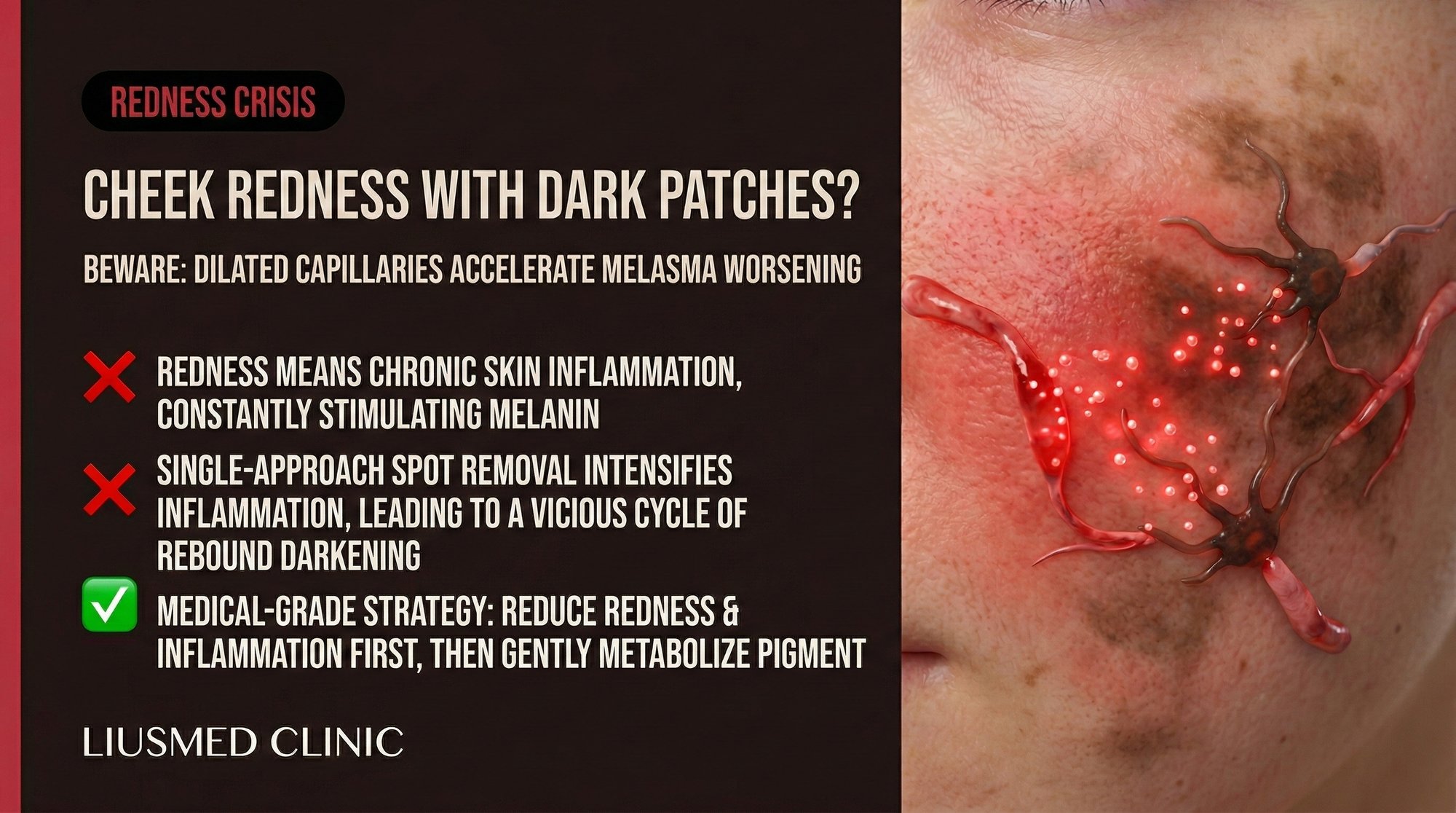
You notice it most in the afternoon or after a warm shower: a frustrating combination of redness and dark patches across your cheeks, as if two completely different skin problems have decided to occupy the same territory. You treat the pigmentation with lightening products. You treat the redness with anti-inflammatory creams. Neither improves significantly, and sometimes one treatment seems to make the other condition worse. This is not a coincidence — when telangiectasia (visible dilated blood vessels) coexists with melasma, the two conditions are not merely neighbours sharing the same real estate. They are active collaborators, each making the other more severe and more resistant to treatment.
Table of Contents
- When Red Meets Brown: The Clinical Overlap
- Telangiectasia and the Melanocyte Connection
- Why Rosacea Patients Are Especially Vulnerable
- The Diagnostic Challenge: Redness Masking Pigmentation
- Treatment Approaches for Combined Vascular-Pigmentary Conditions
- Building a Sequential Treatment Strategy
When Red Meets Brown: The Clinical Overlap
The coexistence of facial redness and hyperpigmentation is far more common than most patients realize. Clinical surveys of melasma patients consistently find that a significant proportion have concurrent telangiectasia, background erythema, or diagnosable rosacea. Conversely, studies of rosacea patients reveal higher-than-expected rates of melasma, particularly in skin types III through V on the Fitzpatrick (Fitzpatrick Skin Type) scale.
This overlap is not random. The cheeks — the most common site for both conditions — represent a vascular watershed zone on the face. The malar region receives blood supply from branches of both the facial artery and the transverse facial artery, creating a rich vascular network close to the skin surface. This anatomical predisposition means that any process affecting dermal blood vessels will be most visible and most impactful in precisely the area where melasma most commonly develops.
Patients with this combined presentation typically report a characteristic progression. Initial symptoms are vascular: intermittent flushing, heat sensitivity, and occasional visible redness. Over months, the redness becomes more persistent, and telangiectasia — fine, visible blood vessels — become apparent. Then pigmentation begins to develop in the same areas, initially subtle but progressively more prominent. By the time the patient seeks treatment, both conditions are well established and intertwined.
The diagnostic challenge is that the redness and pigmentation can obscure each other. Background erythema can make pigmented patches appear less defined, and the brown pigment can mask the full extent of underlying vascular changes. A clinician who focuses only on the pigmentation misses the vascular driver. A clinician who focuses only on the redness misses the opportunity to prevent progressive hyperpigmentation.
Telangiectasia and the Melanocyte Connection
Telangiectasia refers to permanently dilated small blood vessels (capillaries, arterioles, and venules) in the superficial dermis that become visible through the skin surface. In isolation, they cause redness and flushing. But their impact extends far beyond cosmetic redness — they fundamentally alter the microenvironment of the surrounding tissue in ways that directly stimulate melanocyte activity.
Dilated blood vessels release a cocktail of pro-melanogenic factors. Endothelin-1 (ET-1), released by vascular endothelial cells, is one of the most potent known stimulators of melanocyte proliferation and melanin synthesis. Stem cell factor (SCF) from perivascular cells promotes melanocyte survival and differentiation. Basic fibroblast growth factor (bFGF) and hepatocyte growth factor (HGF) from the vascular niche further amplify melanocyte activity.
Beyond these direct paracrine signals, dilated vessels create a pro-inflammatory microenvironment through several mechanisms. Increased blood flow raises local skin temperature, which has been independently shown to stimulate melanogenesis. Greater vascular permeability allows inflammatory mediators — histamine, prostaglandins, leukotrienes — to diffuse more readily into the perivascular dermis and overlying epidermis. Mast cells, which cluster around blood vessels, degranulate more frequently in areas of chronic vascular dilation, releasing additional histamine and VEGF (Vascular Endothelial Growth Factor — new blood vessel signal).
The net result is that each telangiectatic vessel functions as a miniature melanocyte-activating unit. The more vessels present, the greater the surface area of melanocyte stimulation. This creates a direct dose-response relationship: patients with more severe telangiectasia tend to develop more severe and more treatment-resistant melasma.
| Vascular Factor | Source | Effect on Melanocytes | Net Impact on Melasma |
|---|---|---|---|
| Endothelin-1 | Endothelial cells | Increases melanin synthesis and melanocyte proliferation | Strong pro-pigmenting effect |
| VEGF | Endothelial cells, mast cells | Promotes angiogenesis, amplifies vascular component | Self-perpetuating cycle |
| Stem cell factor | Perivascular fibroblasts | Melanocyte survival and differentiation | Maintains melanocyte population |
| Histamine | Mast cells | Increases vascular permeability, indirect melanocyte stimulation | Inflammatory amplification |
| Prostaglandin E2 | Inflammatory cells | Direct melanocyte stimulation via EP receptors | Pigment overproduction |
| Local heat | Increased blood flow | Stimulates tyrosinase activity | Temperature-dependent darkening |
| Reactive oxygen species | Inflammatory infiltrate | DNA damage, melanocyte activation | Chronic oxidative stimulation |
Why Rosacea Patients Are Especially Vulnerable
Rosacea and vascular melasma share a remarkable amount of underlying pathophysiology, making rosacea patients particularly susceptible to developing stubborn, treatment-resistant pigmentation.
Both conditions involve dysfunction of the innate immune system in the skin, with overexpression of cathelicidin (LL-37) and increased toll-like receptor 2 (TLR2) activity. Both show elevated levels of matrix metalloproteinases (MMPs), which degrade the extracellular matrix and basement membrane zone. Both demonstrate mast cell hyperactivity and VEGF overexpression. And both are exacerbated by UV radiation, heat, and emotional stress.
The shared pathway most relevant to pigmentation is the neurovascular connection. Rosacea involves dysregulation of the transient receptor potential vanilloid 1 (TRPV1) channels on sensory nerve endings in the skin. Activation of these channels triggers neurogenic inflammation — the release of substance P, calcitonin gene-related peptide (CGRP), and other neuropeptides that cause vasodilation and inflammatory cell recruitment. These same neuropeptides have been shown to directly stimulate melanocyte activity.
Patients with erythematotelangiectatic rosacea (ETR) — the subtype characterized primarily by persistent redness and visible blood vessels — have the highest risk of developing concurrent vascular melasma. The chronically dilated vascular bed creates exactly the pro-melanogenic microenvironment described in the previous section. Over time, pigmentation develops in the same distribution as the redness, creating the characteristic mixed presentation.
A critical clinical implication is that treating the rosacea component first — reducing the vascular inflammation and stabilizing the telangiectatic vessels — often produces unexpected improvement in the melasma as well, even without specific depigmenting therapy. This observation provides strong indirect evidence that the vascular component is actively driving the pigmentation rather than merely coexisting with it.
The Diagnostic Challenge: Redness Masking Pigmentation
Accurately assessing the relative contributions of vascular redness and melanin pigmentation in the same lesion is genuinely difficult, even for experienced clinicians. The red hemoglobin chromophore and the brown melanin chromophore absorb different wavelengths of light but their visual contributions overlap significantly, making clinical estimation unreliable.
Several diagnostic tools can help disentangle the two components. Dermatoscopy under polarized light preferentially visualizes deeper structures, allowing assessment of the reticular vascular pattern beneath surface pigmentation. Crossed polarization reduces surface reflection and epidermal melanin contribution, making the vascular pattern more apparent.
Wood lamp examination under 365 nm UV light enhances epidermal melanin while being largely transparent to the vascular component. Comparing the clinical appearance under visible light (melanin plus hemoglobin) with the Wood lamp appearance (melanin only) provides an estimate of how much the vascular component contributes to the overall discoloration.
Diascopy — pressing a glass slide against the skin to blanch the blood vessels — is a simple bedside technique that can reveal the true extent of melanin pigmentation by temporarily removing the vascular contribution. If the discoloration largely disappears with diascopy, the dominant component is vascular. If significant brown pigment remains, melanin deposition is confirmed. Most combined cases show partial blanching, indicating both components are present.
The most advanced option is multispectral imaging, which can mathematically separate the melanin and hemoglobin contributions to the visible skin colour. While not widely available, this technology provides the most precise characterization of the relative contributions and is particularly valuable for treatment planning and monitoring.
Understanding the ratio of vascular to pigmentary contribution has direct treatment implications. A predominantly vascular presentation may respond well to anti-redness therapies alone, while a predominantly pigmentary presentation with incidental telangiectasia requires the full melasma treatment arsenal.
Treatment Approaches for Combined Vascular-Pigmentary Conditions
Treating the combined condition requires addressing both the vascular and pigmentary components — ideally in a coordinated sequence that disrupts the feedback loop between them.
Anti-inflammatory foundation. Before attempting depigmentation, the inflammatory environment must be stabilized. This means identifying and avoiding triggers that provoke flushing and vasodilation: extreme temperatures, spicy food, alcohol, aggressive skincare products, and known irritants. A simplified, gentle skincare routine with a focus on barrier repair reduces the background inflammation that drives both conditions.
Vascular-targeted therapy. Reducing the telangiectatic vessel network removes the melanocyte-activating signals at their source. For mild to moderate telangiectasia, topical brimonidine or oxymetazoline can provide temporary vasoconstriction and symptomatic redness relief. For established telangiectasia that requires structural reduction, Melasma Injection Treatment offers a targeted approach that addresses the vascular infrastructure supporting the pigmentary process.
Systemic anti-inflammatory and anti-vascular agents. Oral tranexamic acid serves double duty in combined cases — it reduces plasmin-mediated VEGF production (anti-vascular) and directly suppresses melanocyte activity (anti-pigmentary). Low-dose doxycycline (40 mg modified-release, sub-antimicrobial dose) can address the rosacea-associated inflammation without antibiotic side effects.
Topical depigmenting agents. Once the vascular-inflammatory component is under control, topical depigmenting therapy can be introduced. The sequence matters: starting depigmenting agents before controlling the vascular component often leads to irritation that worsens the redness, which in turn stimulates more pigmentation. Azelaic acid is a particularly elegant choice for combined cases because it has both depigmenting and anti-inflammatory properties at concentrations of 15 to 20 percent.
Photoprotection. Tinted mineral sunscreen with iron oxides is essential. Beyond UV protection, the tinted formulation provides immediate cosmetic camouflage that reduces the visual impact of both redness and pigmentation — a psychological benefit that should not be underestimated for patients living with a visually distressing condition.
Building a Sequential Treatment Strategy
The optimal treatment sequence for combined telangiectasia and melasma follows a logical order: stabilize first, then treat the vascular driver, then address the pigmentation.
Phase 1 (Weeks 1-4): Stabilization. Simplify the skincare routine to cleanser, moisturizer, and tinted mineral sunscreen. Eliminate all active ingredients that could cause irritation. Identify and avoid vasodilatory triggers. Begin oral tranexamic acid if medically appropriate.
Phase 2 (Weeks 4-12): Vascular reduction. Introduce targeted therapy for the telangiectatic component. This is the phase where the vascular infrastructure that sustains the melasma is dismantled. Patients often notice that the pigmentation begins to lighten during this phase even without depigmenting agents — a sign that the vascular contribution to the visible discoloration was substantial.
Phase 3 (Weeks 8-24): Pigment suppression. With the vascular component reduced, introduce topical depigmenting agents. The melanocytes are no longer receiving constant pro-melanogenic signals from dilated vessels, so the topical agents can work as intended without being overwhelmed by vascular stimulation. Azelaic acid, tranexamic acid serum, or controlled-use hydroquinone are appropriate choices.
Phase 4 (Ongoing): Maintenance. Continue tinted mineral sunscreen daily. Maintain trigger avoidance. Monitor for early signs of vascular recurrence, which typically precedes pigmentary relapse. Early intervention at the first sign of increased redness can prevent the full cascade from re-establishing.
This sequential approach is more time-intensive than simply applying a lightening cream, but the outcomes are dramatically more durable because each phase builds on the previous one, and the fundamental vascular driver is addressed rather than ignored.
Frequently Asked Questions
Q1: Can I use anti-redness products and lightening products at the same time?
Using both simultaneously is possible but must be done carefully. The primary risk is over-treating the skin and causing irritation that worsens both conditions. If you plan to use both, introduce them sequentially — establish tolerance to one before adding the other — and choose the gentlest effective formulations. Better results are typically achieved by treating the vascular component first and then adding depigmenting agents once redness is controlled.
Q2: Will laser treatment for my visible blood vessels also help my melasma?
Vascular lasers (such as pulsed dye laser or KTP laser) can effectively treat telangiectasia, and in some cases the melasma improves secondarily as the vascular stimulus is removed. However, the thermal energy from laser treatment carries a risk of triggering post-inflammatory hyperpigmentation, particularly in darker skin types. This risk must be carefully weighed against the potential benefit, and treatment parameters must be conservative.
Q3: I was told I have rosacea, not melasma. Could I have both?
Absolutely. Rosacea and melasma frequently coexist, particularly in women with Fitzpatrick skin types III to V. The two conditions share underlying vascular and inflammatory pathways. If you have been diagnosed with rosacea and also notice brown or grey-brown patches in the same areas as your redness, request evaluation for concurrent melasma. Treatment plans that address only one condition while ignoring the other are often suboptimal.
Q4: Does alcohol really worsen this combined condition?
Yes. Alcohol is a potent vasodilator that causes direct expansion of facial blood vessels. In patients with telangiectasia-associated melasma, alcohol consumption can cause immediate visible darkening of pigmented patches as increased blood flow enhances the hemoglobin contribution and stimulates melanocyte activity. Chronic alcohol use promotes sustained vascular dilation and angiogenesis, worsening both the vascular and pigmentary components over time.
Q5: Is this combined condition more common in certain ethnicities?
The combined presentation is most frequently reported in individuals with moderate skin pigmentation — Fitzpatrick types III through V — who have both the melanocyte reactivity predisposing to melasma and the skin characteristics that make telangiectasia clinically apparent. East Asian, Southeast Asian, Hispanic, and Middle Eastern populations appear to have higher prevalence, though this may partly reflect study population demographics.
Q6: How do I know if my treatment is working when both redness and pigmentation are present?
Standardized photography under consistent lighting conditions is the most reliable method for tracking progress. Ask your clinician to take comparison photos at each visit. You can also perform simple diascopy at home — press a clear glass against the treated area and compare the residual brown pigment over time. As the vascular component improves, you will notice less redness and flushing. As the pigmentary component improves, the residual colour under diascopy will lighten.
About the Author
Dr. Ta-Ju Liu is the founder and lead physician at Liusmed Clinic, specializing in regenerative medicine and minimal incision surgery. His clinical focus on the vascular-inflammatory axis of pigmentary disorders has led to the development of integrated treatment protocols that address both telangiectasia and melasma as components of a unified pathological process. Liusmed Clinic combines precision vascular assessment with targeted intervention therapies for patients with complex, treatment-resistant pigmentation.
Disclaimer
This article is provided for educational and informational purposes only and does not constitute medical advice, diagnosis, or treatment. The coexistence of rosacea, telangiectasia, and melasma creates complex clinical scenarios that require individualized professional assessment. Do not self-diagnose or modify existing treatment plans based on this information alone. Consult a qualified medical professional for evaluation and personalized treatment recommendations. Individual treatment outcomes vary and are not guaranteed.

Specialties
Credentials
- Kaohsiung Medical University, School of Medicine
- Attending Physician, Dermatology, Kaohsiung Chang Gung Memorial Hospital
- Attending Physician, Aesthetic Center, Kaohsiung Chang Gung Memorial Hospital
- Visiting Physician, Dermatology, Xiamen Chang Gung Hospital
- Visiting Physician, Aesthetic Center, Xiamen Chang Gung Hospital
"For every surgery, I strive to achieve a good outcome through a small incision and refined technique. Minimally invasive surgery is not just a technique — it's a commitment of respect to every patient."
Recovery after any procedure needs peer support too
Want to learn more?
Schedule a consultation for professional evaluation and advice