Lump Under Skin? Ultrasound Tells Lipoma from Cyst & Cancer
The sentence I hear most often in clinic is: "Doctor, I felt something while showering — what is it? Should I be worried?"
The moment you feel something under your skin, the anxiety is almost instant. But what unsettles people most is not the lump itself — it is not knowing. Not knowing whether it is benign or malignant, whether to watch it or treat it, and not knowing how large a scar removal would leave.
In the past, answering these questions often meant relying on touch alone, or simply cutting it open to look. Today, high-frequency soft-tissue ultrasound lets us see a subcutaneous lump clearly without any incision at all. This is the heart of Liusmed Clinic's principle — "you can only treat safely what you can see": imaging certainty first, treatment safety second.
This article walks you through what lipomas and epidermoid cysts each look like on ultrasound, when an exam is warranted, how ultrasound helps assess the possibility of malignancy, and how seeing clearly first makes later treatment more precise.
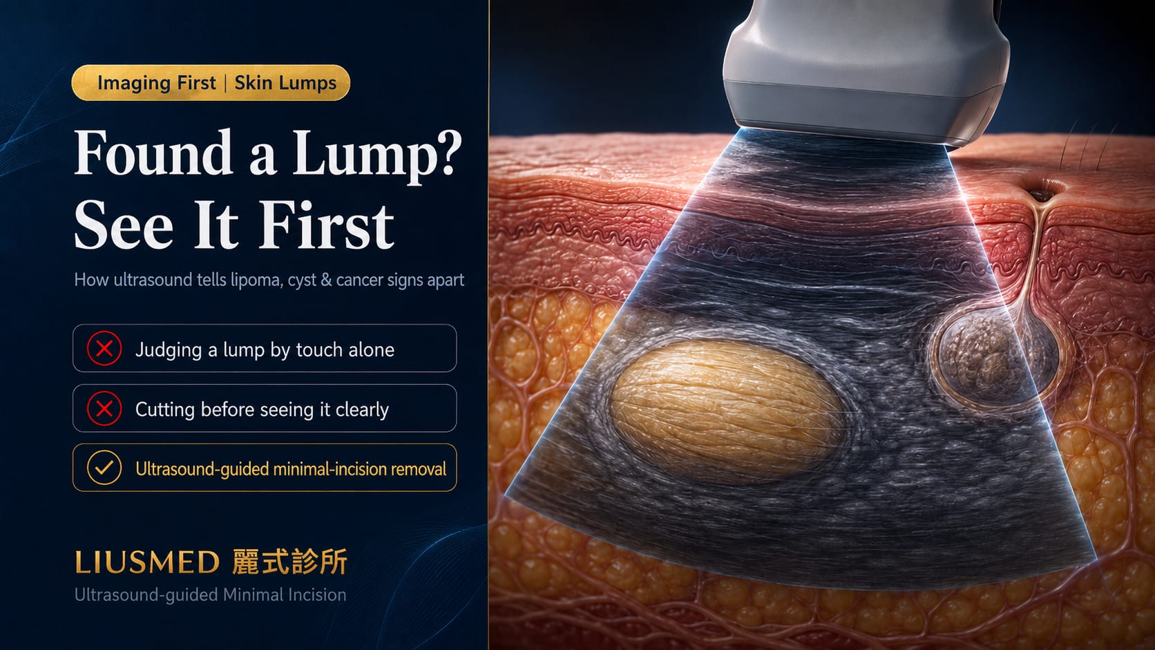
What Lipomas and Cysts Look Like on Ultrasound
A lump under the skin has hundreds of possible causes, but two are by far the most common in clinic: the lipoma (a benign fatty tumor) and the epidermoid cyst (粉瘤). They can feel similar to the touch, yet under ultrasound their imaging features are quite distinct.
Ultrasound works by sending sound waves that reflect differently off different tissues (echoes), then converting those echoes into an image. Tissues differ in firmness, water content and structural arrangement, so they differ in "echogenicity" — and that is exactly what lets us tell lump types apart.
Ultrasound Features of a Lipoma
A lipoma is a benign tumor formed by overgrowth of fat cells. On ultrasound it usually:
- Sits within the subcutaneous fat layer, as an oval with its long axis parallel to the skin
- Has echogenicity close to surrounding fat (isoechoic), often with fine striations parallel to the long axis
- Has clear borders, though not always a complete capsule
- Deforms slightly when pressed with the probe (soft texture)
- Usually shows little blood flow on Doppler
Ultrasound Features of an Epidermoid Cyst
An epidermoid cyst is a sac formed by keratin and sebum accumulating under the skin. Its ultrasound features are quite recognizable:
- Sits more superficially, right against the dermis, often with a thin tract leading to a surface opening
- Shows uniform "pseudo-solid" internal echoes, with posterior acoustic enhancement (the image brightens behind the sac)
- Has clear borders and a defined cyst wall
- When inflamed, the surrounding tissue echoes become disorganized and blood flow increases
| Comparison | Lipoma | Epidermoid cyst |
|---|---|---|
| Tissue layer | Subcutaneous fat, deeper | Against the dermis, more superficial |
| Link to skin surface | None | Often a tract to a surface opening |
| Internal echoes | Isoechoic + parallel striations | Uniform pseudo-solid |
| Posterior echoes | No clear change | Enhanced |
| Deformation on pressure | Slightly deformable | Less deformable |
| Tendency to inflame | Low | Common — can redden and suppurate |
Key point: "Feels like" is not "is." Lipomas and epidermoid cysts differ in how they are treated, their recurrence risk and their tendency to inflame — ultrasound separates the two before any cut, so the wrong approach is not used. To understand the difference in more depth, see the difference between lipoma and cyst.
When a Subcutaneous Lump Should Be Scanned
Not every lump needs imaging. A lump that has been small, soft, mobile and unchanged for years can often be observed after a physical examination. But the following situations warrant an ultrasound to clarify:
- Noticeable growth in a short time — enlarging over weeks to months
- Deep position or unclear borders — physical exam cannot define its extent
- A larger lump, especially one over 5 cm
- Fixed, barely mobile, or attached to deeper tissue
- Tenderness, redness or repeated inflammation
- Before surgery — to know the lump's exact depth, size and relationship to vessels and nerves
- A lump that causes you ongoing anxiety — being certain what it is, is itself part of treatment
Ultrasound has no radiation and is non-invasive, so treating it as "the first confirmation before doing anything" is reasonable. Many people jump straight to "should I cut it out?" — but the question that comes first is what it actually is, and how deep it goes. With answers in hand, every later decision rests on firmer ground.
Can Ultrasound Reveal Warning Signs of Malignancy
This is what patients care about most — and the point that most needs an honest answer.
The overwhelming majority of subcutaneous lumps are benign. But one malignant tumor — the liposarcoma — can look similar to a benign lipoma in its early stages and is easily dismissed. Here, ultrasound's value is helping flag the signs that "do not look like a simple lipoma":
- Large size (often over 5 cm) and continued growth
- Uneven internal echoes, with nodular or solid-looking regions
- Irregular borders, infiltrating into surrounding tissue
- Markedly increased blood flow on Doppler
- Deep location, such as within a muscle layer
When these features appear, it means "further investigation is needed" — not "remove it in clinic right away."
Key point: Ultrasound is an excellent screening and differentiation tool, but it cannot rule out malignancy with 100% certainty. Deep, rapidly enlarging or imaging-suspicious lumps still need MRI (Magnetic Resonance Imaging) or a pathology biopsy to confirm. If your lump matches any of the warning signs above, the correct next step is assessment by a specialist — not self-judgment or delay.
For this reason, at Liusmed Clinic every subcutaneous tumor procedure is built on "seeing clearly first" — minimal-incision removal proceeds only when imaging confirms a benign, well-defined lump; when imaging raises doubt, further investigation comes first.
What the Exam Is Like and What to Note
Many people have never had a soft-tissue ultrasound and imagine it as more complex than it is. The process is actually straightforward:
- No special preparation — no fasting, no stopping medication; your normal routine is fine
- Coupling gel is applied to the skin, and the doctor scans the lump area with a high-frequency probe
- The process is painless and radiation-free, with the image visible in real time
- The doctor measures the lump's size, depth, tissue layer and blood flow on the spot, and explains the findings to you
- The whole exam takes about 10 to 15 minutes
The one thing worth noting: interpreting a soft-tissue ultrasound depends heavily on operator experience. On the same machine, the detail two doctors see can differ greatly. Having the same doctor who performs the tumor removal also scan and interpret the image removes any information gap between "the exam" and "the treatment" — what is seen is handled by the one who saw it.
From "Seeing" to "Treating Safely": Ultrasound-Guided Removal
Ultrasound's value lies not only in diagnosis but extends into treatment.
Traditionally, subcutaneous tumor removal relied largely on feel and experience, and the wound often had to be made larger than the lump to extract it completely. But when the doctor can use ultrasound to precisely locate the lump's borders, depth and relationship to vessels and nerves — before and even during surgery — the logic reverses: the lump can be removed completely through the smallest possible wound.
This is Liusmed Clinic's <20% extreme minimal-incision principle: keeping the wound length within 20% of the lesion's diameter. A lump several centimeters across may need only an incision of a fraction of a centimeter to just over one. What makes this possible is, precisely, being able to see.
- For an overview of how subcutaneous tumors are handled, see the skin tumor overview
- For minimal-incision removal of lipomas, see lipoma minimal-incision surgery and the complete guide to minimal-incision lipoma surgery
- For cyst treatment and complete sac removal, see cyst removal and the complete guide to epidermoid cysts
Frequently Asked Questions
Q1: If I feel a lump under my skin, do I always need an ultrasound?
A1: Not always. A small, soft, mobile lump that has not changed for a long time can often be observed after a physical examination. But if the lump grows quickly, has unclear borders, sits deep, is tender or repeatedly inflamed, or if you are preparing for surgery, ultrasound provides information a physical exam cannot — and is then recommended.
Q2: Does the ultrasound exam hurt? What do I need to prepare?
A2: It does not hurt, and there is no radiation. The exam only involves applying gel to the skin and scanning with a probe, and takes about 10 to 15 minutes. No fasting and no stopping medication are needed — you can come in with your normal routine.
Q3: Can ultrasound confirm whether a lump is benign or malignant?
A3: Ultrasound can effectively distinguish common benign lumps such as lipomas and epidermoid cysts, and can also flag signs that "do not look benign." But it cannot rule out malignancy with 100% certainty — imaging-suspicious, large or deep lumps still need MRI or a pathology biopsy to confirm. Treat ultrasound as an important first screening step, not a final diagnosis.
Q4: If the exam shows it is benign, can it be removed right away?
A4: A lump confirmed on imaging to be benign and well-defined can usually be scheduled for minimal-incision removal in clinic. The depth and border information ultrasound provides is exactly what allows the incision to be kept as small as possible. The actual approach is still decided by the doctor based on the lump's location, size and your needs.
See It Clearly First, Then Decide How to Treat It
The anxiety of feeling a lump under your skin often comes from "not knowing." And the most direct way to remove "not knowing" is to let the lump be seen.
A subcutaneous tumor ultrasound exam is painless and radiation-free, yet before any cut it lays out the lump's type, depth, borders and risk signals. For benign lumps, it makes later minimal-incision removal more precise; for the few suspicious lesions, it gives early warning that further investigation is needed.
Liusmed Clinic director Dr. Ta-Ju Liu has long focused on the minimal-incision management of subcutaneous tumors, insisting that every procedure rests on "seeing clearly first." If a lump you have found is troubling you, you are welcome to book a consultation — let us see it clearly together first, then decide on the next step.

Specialties
Credentials
- Kaohsiung Medical University, School of Medicine
- Attending Physician, Dermatology, Kaohsiung Chang Gung Memorial Hospital
- Attending Physician, Aesthetic Center, Kaohsiung Chang Gung Memorial Hospital
- Visiting Physician, Dermatology, Xiamen Chang Gung Hospital
- Visiting Physician, Aesthetic Center, Xiamen Chang Gung Hospital
"For every surgery, I strive to achieve a good outcome through a small incision and refined technique. Minimally invasive surgery is not just a technique — it's a commitment of respect to every patient."
Want to learn more?
Schedule a consultation for professional evaluation and advice