Symmetrical Reticular Dark Patches on Your Face? Understanding the Most Stubborn Vascular Melasma
You have tried every lightening product available. You have been meticulous with sunscreen. Yet the brown patches on your cheeks persist — and when you look closely, the discoloration is not a solid block of colour but a web-like, net-patterned stain that seems to sit deeper than ordinary pigmentation. If this describes your experience, you are likely dealing with vascular melasma — a subtype that accounts for a significant proportion of treatment-resistant cases and one that most conventional approaches fail to address because they target the wrong layer of the problem.
Table of Contents
- What Makes Vascular Melasma Different
- The Reticular Pattern: Reading the Map on Your Skin
- The Blood Vessel–Melanocyte Feedback Loop
- Why Standard Melasma Treatments Fall Short
- Diagnostic Methods for Identifying Vascular Melasma
- Evidence-Based Approaches for Vascular Melasma
What Makes Vascular Melasma Different
Melasma is not a single disease — it is an umbrella term for a spectrum of pigmentary conditions that share a common clinical appearance but differ substantially in their underlying pathophysiology. The traditional classification divides melasma into epidermal, dermal, and mixed subtypes based on pigment depth. But this framework misses an entire dimension of the disease: the vascular component.
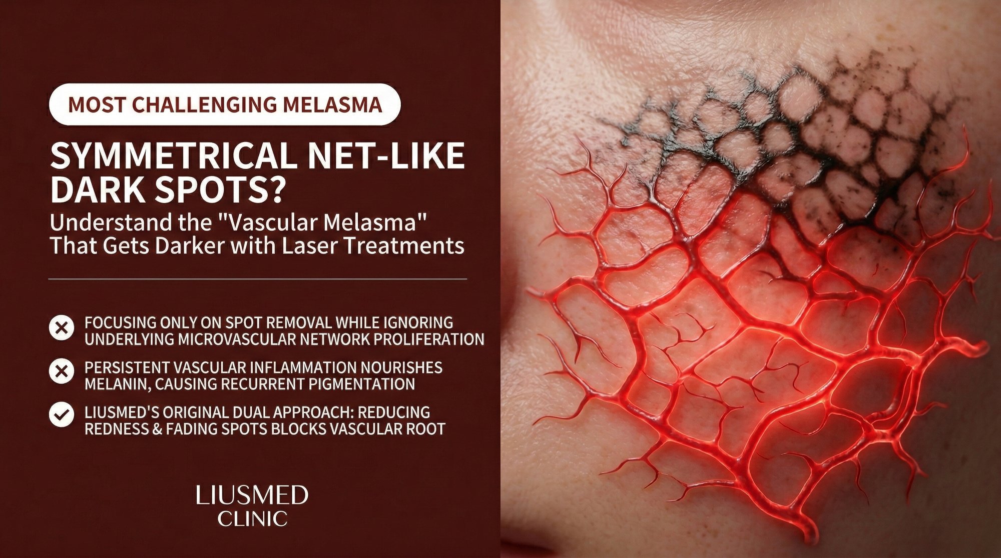
Vascular melasma is characterized by the presence of an abnormally dense network of dilated blood vessels — telangiectasia and neovascular channels — in the dermis directly beneath the pigmented patches. These are not incidental findings. Histological studies comparing melasma-affected skin to adjacent normal skin in the same patient consistently demonstrate significantly increased vascular density, larger vessel calibre, and elevated expression of vascular endothelial growth factor (VEGF) within the melasma lesion.
The practical consequence is straightforward: these blood vessels actively maintain the conditions that keep melanocytes overproducing pigment. You can bleach the melanin away with hydroquinone, destroy it with a laser, or suppress it with tranexamic acid — but as long as the vascular infrastructure beneath remains intact, the melanocytes receive a constant stream of growth factors and inflammatory signals that drive repigmentation within weeks to months.
This is the fundamental reason why so many melasma patients experience the demoralizing cycle of initial improvement followed by inevitable relapse. The treatment addressed the symptom (excess melanin) without touching the cause (vascular-mediated melanocyte activation).
The Reticular Pattern: Reading the Map on Your Skin
The reticular pattern — a net-like or mesh-like distribution of pigment — is the visible signature of vascular melasma. Unlike the homogeneous brown patches seen in purely epidermal melasma, vascular melasma produces a pattern where darker lines of pigment follow the branching architecture of the underlying blood vessels, creating a lattice of discoloration interspersed with relatively lighter areas.
This pattern is most clearly visible under dermatoscopic examination, where it is classified as a reticular or pseudo-reticular network. Under the Wood lamp, vascular melasma often shows less enhancement than epidermal melasma — the pigment does not glow as brightly because a significant component of the visible discoloration comes from the hemoglobin in the dilated vessels rather than from melanin alone.
Several clinical features can help identify this pattern even without specialized instruments. The patches often have a slightly warm or reddish undertone rather than the cool grey-brown of pure dermal melasma. The borders may be less defined than in epidermal melasma, with the pigment appearing to diffuse outward like watercolour rather than having clear demarcation. And the pigment intensity fluctuates more with vasodilatory triggers — heat, alcohol, emotional stress, exercise — than it does with UV exposure alone.
Patients with vascular melasma frequently report a characteristic progression: they notice faint redness in an area first, followed weeks or months later by progressive darkening of the same zone. This timeline makes biological sense — the vascular changes establish the inflammatory microenvironment first, and the melanocyte response follows as the cells adapt to the chronic stimulation.
The Blood Vessel–Melanocyte Feedback Loop
Understanding vascular melasma requires understanding a critical biological concept: the paracrine feedback loop between dermal blood vessels and epidermal melanocytes. In normal skin, these two cell populations communicate at low levels — blood vessels supply nutrients and carry away waste, while melanocytes respond to local signals to maintain appropriate pigmentation. In vascular melasma, this communication becomes pathologically amplified.
The cycle begins with vascular activation. Whether triggered by UV radiation, hormonal changes, inflammation, or a combination of factors, blood vessels in the affected dermis begin to dilate and proliferate. These activated vessels release VEGF (Vascular Endothelial Growth Factor — new blood vessel signal), which promotes further angiogenesis — more blood vessel growth. They also release endothelin-1, stem cell factor, basic fibroblast growth factor, and various inflammatory cytokines. Each of these molecules can independently stimulate melanocyte activity.
Melanocytes respond to this flood of signals by increasing tyrosinase expression and melanin production. But melanocytes are not passive recipients — they also release factors that further promote vascular growth, including VEGF itself. The result is a self-sustaining cycle: vessels stimulate melanocytes, melanocytes stimulate vessels, and both populations maintain each other in a state of chronic overactivity.
Additional amplifying factors include mast cells, which are consistently found in higher numbers in melasma-affected dermis. Mast cells release histamine, tryptase, and other mediators that promote both vasodilation and melanogenesis. They also release VEGF, adding another arm to the feed-forward cycle. This mast cell involvement may explain why antihistamines have shown modest benefit in some melasma cases — not enough to serve as standalone treatment, but enough to suggest that the inflammatory-vascular axis is therapeutically relevant.
The depth of this feedback loop explains the characteristic treatment resistance of vascular melasma. Any therapy that targets only one element of the cycle — melanin, melanocyte activity, inflammation, or vasculature — provides temporary benefit at best. The untreated elements continue to operate, and the cycle re-establishes itself as soon as the suppressive therapy is discontinued.
Why Standard Melasma Treatments Fall Short
The standard melasma treatment algorithm — topical depigmenting agents, sun protection, and sometimes chemical peels or gentle laser toning — was developed primarily based on the epidermal melasma model. For vascular melasma, each of these approaches has specific limitations.
| Treatment | Mechanism | Why It Fails in Vascular Melasma |
|---|---|---|
| Hydroquinone | Inhibits tyrosinase enzyme | Does not affect vascular proliferation or inflammatory mediators; melanocytes are re-stimulated by vascular signals after discontinuation |
| Tretinoin | Accelerates epidermal turnover | Epidermal effects only; no impact on dermal vasculature; can cause irritation that worsens vascular component |
| Vitamin C | Antioxidant, mild tyrosinase inhibitor | Insufficient concentration reaches dermal vessels; antioxidant effect too weak to break the feedback loop |
| Chemical peels | Remove superficial pigmented cells | Epidermal effect only; inflammatory response from peel can stimulate vascular component |
| Laser toning | Fragment melanin particles | Thermal energy can dilate vessels and trigger inflammation; risk of paradoxical darkening in vascular subtype |
| IPL | Target melanin and hemoglobin | May partially address vascular component but risk of post-inflammatory hyperpigmentation; insufficient vessel-specific targeting |
| Oral tranexamic acid | Anti-plasmin, reduces VEGF | Partially effective — addresses some vascular signaling but may not be sufficient as monotherapy for severe vascular melasma |
The pattern is clear: treatments that work well for epidermal melasma systematically underperform in vascular melasma because they ignore the vascular-inflammatory infrastructure that sustains the condition. The few therapies that do address the vascular component — oral tranexamic acid being the most studied — show better results, but even these are often insufficient as standalone interventions for moderate to severe vascular melasma.
Diagnostic Methods for Identifying Vascular Melasma
Accurate identification of the vascular component is essential before treatment planning. Several diagnostic tools can help determine whether your melasma has significant vascular involvement.
Dermatoscopy is the most accessible and informative tool. A trained dermatoscopist can identify the reticular vascular pattern, assess the relative contributions of melanin and hemoglobin to the visible pigmentation, and evaluate the density and calibre of visible telangiectasia within the lesion. Polarized dermatoscopy is particularly useful for visualizing deeper vascular structures.
Wood lamp examination provides complementary information. Purely epidermal melanin enhances under Wood lamp illumination, making the lesion boundaries more prominent. Vascular melasma shows less enhancement because the hemoglobin component does not fluoresce. A lesion that is clearly visible to the naked eye but shows only modest enhancement under Wood lamp should raise suspicion for a significant vascular or dermal component.
Reflectance confocal microscopy (RCM) represents the gold standard for non-invasive melasma characterization. RCM can visualize melanin distribution at the cellular level and identify dilated blood vessels in the superficial dermis in real time. However, this technology is not widely available outside specialized dermatology centres.
Clinical history remains an invaluable diagnostic tool. Ask yourself: Does the pigmentation worsen with heat, alcohol, spicy food, or emotional stress? Does the area show a reddish undertone? Has the condition been resistant to conventional topical depigmenting therapy for more than six months? Does the pigment appear to have a web-like rather than solid pattern? If the answer to two or more of these questions is yes, vascular melasma is highly likely.
Evidence-Based Approaches for Vascular Melasma
Effective management of vascular melasma requires a multi-target strategy that addresses both the pigmentary and vascular components simultaneously. The treatment plan should be sequenced so that the vascular infrastructure is disrupted first or concurrently with melanin suppression — otherwise the melanin will simply return.
The foundation remains rigorous photoprotection with tinted mineral sunscreen containing iron oxides for visible light protection. This is non-negotiable regardless of the melasma subtype. However, for vascular melasma, additional environmental trigger avoidance is important: minimize exposure to extreme heat, limit alcohol consumption, and manage emotional stress where possible.
Oral tranexamic acid has the strongest evidence base for vascular melasma among systemic medications. At typical doses of 250 mg twice daily, it reduces plasmin activity, which in turn reduces VEGF production and prostaglandin synthesis. Clinical trials consistently show superior outcomes with oral tranexamic acid compared to topical therapy alone for the vascular subtype.
For patients with moderate to severe vascular melasma who have not responded adequately to topical and oral pharmacotherapy, targeted vascular interventions become the next logical step. Melasma Injection Treatment at Liusmed Clinic addresses the vascular component directly by targeting the abnormal vessel network that sustains the melanocyte feedback loop. By reducing the vascular infrastructure, the inflammatory signals that drive chronic melanin overproduction are interrupted at their source.
Maintenance therapy is essential regardless of the treatment modality used. Vascular melasma has a high recurrence rate because the factors that originally promoted vascular proliferation — hormonal fluctuations, UV exposure, genetic predisposition — do not disappear. Long-term management typically involves ongoing photoprotection, periodic assessment of the vascular component, and early intervention at the first signs of recurrence.
Frequently Asked Questions
Q1: How can I tell if my melasma is vascular or purely pigmentary?
The most reliable indicator is the pattern and behaviour of the pigmentation. Vascular melasma tends to show a reticular or net-like pattern, has a reddish undertone, worsens with heat and vasodilatory triggers, and responds poorly to conventional lightening agents. A dermatoscopic examination by a trained physician can confirm the presence of abnormal vasculature beneath the pigmented area.
Q2: Is vascular melasma more common than other subtypes?
Recent studies using advanced imaging suggest that the vascular component is present in a larger proportion of melasma cases than previously recognized — potentially the majority. Many cases classified as "treatment-resistant mixed melasma" likely have a significant unrecognized vascular component that explains their resistance to standard therapy.
Q3: Can vascular melasma develop from repeated aggressive laser treatments?
Yes. Aggressive laser treatments that cause significant thermal injury to the skin can promote inflammatory angiogenesis — the growth of new blood vessels in response to tissue damage. This is one reason why melasma can paradoxically worsen after laser therapy. The thermal injury creates exactly the vascular-inflammatory environment that perpetuates vascular melasma.
Q4: Does rosacea increase the risk of vascular melasma?
There is a strong clinical association between rosacea and vascular melasma, likely because both conditions share underlying pathophysiology related to vascular dysregulation, chronic inflammation, and mast cell activation. Patients with concurrent rosacea and melasma often find that treating the rosacea first — reducing baseline vascular inflammation — improves the melasma as well.
Q5: Will the blood vessels in vascular melasma go away on their own?
In most cases, no. Once established, the neovascular network in vascular melasma is self-sustaining due to the feedback loop between vessels, melanocytes, and inflammatory cells. Removing the initial trigger (such as discontinuing hormonal contraceptives) may slow the progression but is unlikely to cause regression of established vasculature. Active treatment targeting the blood vessels is typically required.
Q6: How long does treatment for vascular melasma take compared to epidermal melasma?
Vascular melasma generally requires a longer treatment timeline. While epidermal melasma may show significant improvement within 8 to 12 weeks, vascular melasma often requires 4 to 6 months of multi-target therapy before meaningful improvement is visible. The vascular infrastructure takes time to remodel, and the melanocytes need a sustained period without vascular stimulation before they return to normal baseline activity.
About the Author
Dr. Ta-Ju Liu is the founder and lead physician at Liusmed Clinic, specializing in regenerative medicine and minimal incision surgery. His approach to pigmentary disorders integrates vascular assessment and targeted intervention, reflecting the current understanding that treatment-resistant melasma is fundamentally a vascular-inflammatory condition. Liusmed Clinic employs advanced diagnostic tools and multi-target treatment protocols designed to address the root vascular pathology of stubborn melasma.
Disclaimer
This article is intended for educational and informational purposes only and does not constitute medical advice, diagnosis, or treatment. Vascular melasma is a complex condition that requires individualized professional assessment. The information provided here reflects current medical understanding but may not apply to every case. Consult a qualified dermatologist or medical professional before beginning any treatment. Individual results vary and are not guaranteed.

Specialties
Credentials
- Kaohsiung Medical University, School of Medicine
- Attending Physician, Dermatology, Kaohsiung Chang Gung Memorial Hospital
- Attending Physician, Aesthetic Center, Kaohsiung Chang Gung Memorial Hospital
- Visiting Physician, Dermatology, Xiamen Chang Gung Hospital
- Visiting Physician, Aesthetic Center, Xiamen Chang Gung Hospital
"For every surgery, I strive to achieve a good outcome through a small incision and refined technique. Minimally invasive surgery is not just a technique — it's a commitment of respect to every patient."
Recovery after any procedure needs peer support too
Want to learn more?
Schedule a consultation for professional evaluation and advice