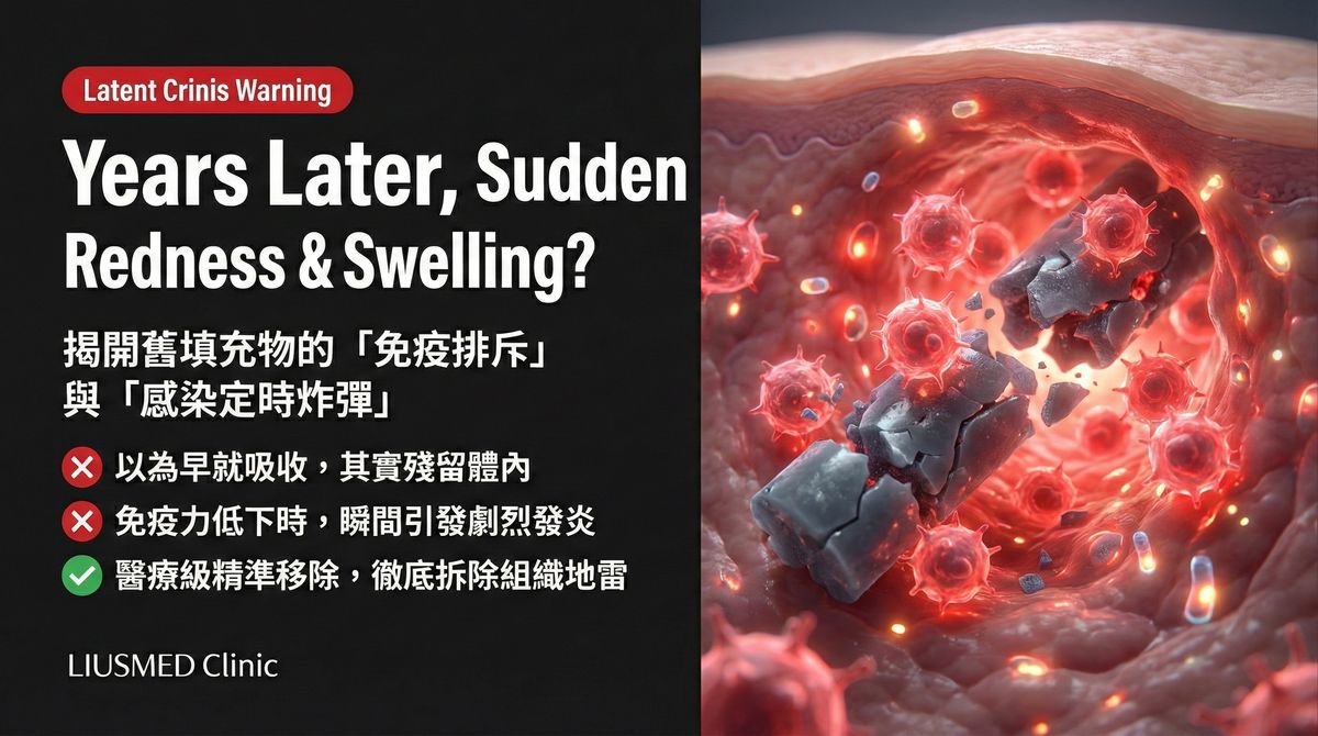
Sudden Redness and Swelling Years After Filler? Immune Rejection and Infection Risks Explained
Filler Injected Years Ago—Why Is It Suddenly Swollen?
Three years ago you had filler in your nasolabial folds. Or five years ago you augmented your cheeks. Everything was uneventful—until one day you look in the mirror and see that area suddenly red and swollen. It feels warm, perhaps even painful. You think in alarm: "How can something I had done years ago suddenly cause problems now?"
This scenario is more common than you might think. Filler-related delayed reactions can first appear months, years, or even more than a decade after injection. Understanding the mechanisms is the first step toward correct management.
Why Did It Stay "Quiet" for So Long Before Erupting?
Three Possible Mechanisms
Sudden redness and swelling years later is usually driven by one of three mechanisms (or their combination):
| Mechanism | Trigger Pattern | Typical Timeframe | Prognosis |
|---|---|---|---|
| Biofilm reactivation | Immune balance disruption | Months to decades post-injection | Requires physical removal of filler |
| Immune-mediated foreign body reaction | Filler degradation or surface change | 1–10 years post-injection | Depends on severity |
| Degradation product reaction | Material breakdown releasing fragments | 2–10 years post-injection | Depends on material type |
Mechanism 1: Biofilm Reactivation
The Sleeping Enemy Awakens
This is the most common cause. As discussed in our biofilm article, bacteria may attach to the filler surface on the day of injection, forming a protected dormant colony. Normally, your immune system and the biofilm maintain a delicate equilibrium—bacteria remain inactive, the immune system maintains low-level surveillance.
But this balance can be disrupted:
- Systemic infection: Influenza, COVID-19, pneumonia temporarily redirecting immune resources
- Immunosuppression: Immunosuppressive medications, chronic stress, malnutrition
- Vaccination: Vaccine-induced systemic immune activation causing cross-reactivity at biofilm sites
- Local trauma: Facial impact, surgery, or other treatments disturbing the local environment
- Hormonal changes: Pregnancy, menopause, or hormonal therapies altering immune function
Key Insight: Biofilm reactivation is not a "new infection"—it is an infection that has existed since injection day, becoming active again when conditions change. This is why antibiotics only temporarily control symptoms and cannot cure the condition. The definitive solution is physical removal of the biofilm-harboring filler.
How to Recognize Biofilm Reactivation
Biofilm reactivation has characteristic features:
- Recurrent episodes—swelling resolves then returns
- Antibiotics provide temporary relief but symptoms recur after discontinuation
- Swelling correlates with overall health status
- Swelling location corresponds to the original injection site
Mechanism 2: Immune-Mediated Foreign Body Reaction
Your Immune System Finally "Sees" the Filler
Even the most biocompatible filler material remains foreign to the human body. In most cases, the immune system develops "immune tolerance"—acknowledging the filler's presence without attacking it.
But this tolerance may collapse years later:
- Filler surface changes: Over time, protein deposition on the filler surface alters its immunological profile
- Filler fragmentation: As filler begins to degrade, new antigenic surfaces are exposed
- Immune system changes: Autoimmune disease flare, new allergen exposure, or altered immune function
- Cross-reactivity: Infection or vaccine-induced immune responses inadvertently targeting filler
Foreign body reaction differs from biofilm:
- Typically presents as diffuse, uniform swelling rather than focal nodules
- May be accompanied by systemic allergic symptoms (rash, itching)
- Antibiotics are completely ineffective
- Corticosteroids may temporarily help but carry long-term side effects
Mechanism 3: Degradation Product Reaction
Breakdown Fragments Triggering New Problems
Different filler materials degrade through different pathways, producing different fragments:
| Filler Type | Degradation Mode | Fragment Characteristics | Reaction Risk |
|---|---|---|---|
| Hyaluronic acid (HA) | Enzymatic degradation | Small polysaccharide molecules | Lower |
| Poly-L-lactic acid (PLLA) | Hydrolytic degradation | Lactic acid molecules | Moderate |
| Polycaprolactone (PCL) | Slow hydrolysis | Caprolactone fragments | Moderate |
| Calcium hydroxylapatite (CaHA) | Phagocytic degradation | Calcium phosphate particles | Moderate-high |
| PMMA/Silicone | Non-degradable | Not applicable | Persistent foreign body reaction |
Some fillers release microparticles or chemical byproducts during degradation that trigger new immune responses. This commonly occurs 2–5 years post-injection—when the filler enters its active degradation phase.
Key Insight: "Degradable" does not mean "safely disappears." The degradation process itself can be a source of complications, particularly when degradation products provoke excessive immune reactions. See does hyaluronic acid truly get completely absorbed?
The Correct Management Process
Step 1: Do Not Panic, But Do Not Wait
Sudden redness and swelling is understandably concerning, but in most cases it can be controlled with proper management. What you should do:
- Document symptoms: Photograph the extent, color, and temporal changes of swelling
- Recall triggering events: Recent illness, vaccination, major stress, or health changes
- Recall injection history: When, what material, and where it was injected
- Schedule evaluation: Arrange prompt assessment with a physician equipped with ultrasound
Step 2: Ultrasound Assessment
Ultrasound plays an irreplaceable role in this setting:
- Confirm whether filler remains in its original position—or has migrated
- Assess for fluid collection (abscess or effusion)
- Evaluate the degree and extent of surrounding tissue inflammation
- Check for capsule formation
- Exclude other possible diagnoses
Step 3: Strategy Based on Diagnosis
Different causes require entirely different treatments:
Biofilm infection:
- Short-term: Appropriate antibiotics to control acute symptoms
- Definitive: Ultrasound-guided physical removal of biofilm-harboring filler
Immune-mediated reaction:
- Mild: Immunomodulatory treatment, observation
- Moderate: May require local corticosteroid injection
- Severe: Consider filler removal
Degradation product reaction:
- Assess amount and condition of residual filler
- Remove residual material if needed to eliminate the reaction source
What You Should NOT Do
Common Mistakes
- Self-medicating with antibiotics: Taking antibiotics without physician evaluation is not only potentially ineffective (if it is not infection) but may mask the real problem
- Blind hyaluronidase injection "to see if it helps": Without ultrasound, you do not know where the problem is or whether hyaluronidase can reach it
- Hot compresses: If the cause is infection, heat accelerates bacterial activity and inflammation
- Massage: If filler has migrated or an abscess is present, massage only spreads the problem
- Ignoring it: "It swelled before and resolved on its own"—each recurrence may be worse than the last
Risk Comparison by Filler Material
Not all fillers carry equal delayed reaction risk. Understanding your injected material helps assess your risk level:
| Material | Delayed Reaction Risk | Removability | Notes |
|---|---|---|---|
| Hyaluronic acid | Low–moderate | Hyaluronidase + physical extraction | Most common, but not zero risk |
| Poly-L-lactic acid | Moderate | Physical extraction only | Degradation phase may trigger reactions |
| Polycaprolactone | Moderate | Physical extraction only | Long-lasting but not permanent |
| Calcium hydroxylapatite | Moderate–high | Physical extraction only | Calcification increases removal difficulty |
| PMMA | High | Physical extraction only, difficult | Permanent material, reactions may persist |
| Silicone | High | Extremely difficult | May integrate with tissue |
Key Insight: A material's "longevity" and "safety" are not the same thing. Longer-lasting fillers mean longer foreign body exposure and a wider window for complications. See lumps found years after injection.
Common questions
Why is filler I had years ago only swelling up now?
Because delayed reactions can genuinely surface for the first time months or years after the injection. It is usually one of three things: a dormant biofilm becoming active again, your immune system starting to react to the filler as a foreign body, or the filler's breakdown products setting off inflammation. Often something recent — an illness, a vaccine, a stretch of heavy stress — tips the balance that had been holding.
Will antibiotics just fix it?
If it is a biofilm, antibiotics can quiet the symptoms for a while, but they do not clear the source, so it tends to flare again once you stop. And if it is not an infection at all but an immune reaction, antibiotics do nothing. That is why we do not keep handing them out before working out what is actually going on.
Can I just have it dissolved with hyaluronidase?
Not blindly. Without ultrasound first, you do not know where the filler sits, whether it is even hyaluronic acid, or whether the enzyme can reach it. Injecting on a guess can miss the problem entirely. We look with ultrasound, then decide.
Can I use a hot compress or massage it while it is swollen?
Better not, until you know the cause. If it is an infection, heat can speed the inflammation up; if the filler has migrated or there is a pocket of fluid, massage just spreads it. It feels like you are helping, but it can make things worse.
Why is ultrasound so important here?
Because it lets us actually see what is going on — whether the filler is still in place or has moved, whether fluid is collecting, how inflamed the surrounding tissue is, and whether a capsule has formed. The different causes need completely different handling, and you can only aim the treatment properly once you can see the problem.
A Message for Those Facing This Suddenly
You may be experiencing panic—a treatment you thought was long behind you has suddenly struck years later. I understand the shock and frustration.
But I also want you to know: you are not alone. Delayed reactions are not rare in clinical practice, and the vast majority of cases, when correctly diagnosed and managed, achieve satisfying improvement.
The key is not to guess on your own and not to delay. Find a physician who can "see" your problem with ultrasound. When you can see it, you can treat it.
Schedule a consultation and let us face this challenge together.
Related Reading
- Biofilm: Why Your Filler Keeps Swelling Years Later
- Found a Lump Years After Injection? Do Not Rush to Dissolve
- Why Dissolvers Fail: The Encapsulation Problem
Related Services

Specialties
Credentials
- Kaohsiung Medical University, School of Medicine
- Attending Physician, Dermatology, Kaohsiung Chang Gung Memorial Hospital
- Attending Physician, Aesthetic Center, Kaohsiung Chang Gung Memorial Hospital
- Visiting Physician, Dermatology, Xiamen Chang Gung Hospital
- Visiting Physician, Aesthetic Center, Xiamen Chang Gung Hospital
"For every surgery, I strive to achieve a good outcome through a small incision and refined technique. Minimally invasive surgery is not just a technique — it's a commitment of respect to every patient."
Recovery after any procedure needs peer support too
Want to learn more?
Schedule a consultation for professional evaluation and advice