Permanent Filler Complications (Silicone, PAAG): Avoiding Extensive Surgical Excision
Why Are Decades-Old Cosmetic Injections Causing Problems Now?
Across Taiwan, mainland China, and Southeast Asia, the 1990s through the early 2000s saw widespread use of injectable cosmetic procedures sometimes called "small needle beautification." The materials used during that era included liquid silicone, polyacrylamide hydrogel (PAAG, marketed as Amazingel or Aquamid), paraffin wax, and various other permanent or semi-permanent fillers. For what these permanent materials like silicone and PAAG actually are, the filler-revision site has a permanent / silicone material encyclopedia page.
These substances were injected into the face, nose, chin, forehead, and even the chest, marketed as long-lasting beauty solutions. Decades later, however, a growing number of patients are confronting serious complications: recurrent inflammation, filler migration, granuloma formation, and in severe cases, tissue necrosis.
Key Insight: Permanent filler problems do not "fade with time" — quite the opposite. As tissues age, gravity takes effect, and the immune system fluctuates, complications from these materials tend to worsen progressively.
Common Permanent Fillers and Their Risks
Material Comparison
| Material | Chemical Composition | Era of Use | Primary Risks | Dissolvable? |
|---|---|---|---|---|
| Liquid silicone | Polydimethylsiloxane | 1960s–2000s | Migration, granuloma, chronic inflammation | No |
| PAAG (Amazingel) | Polyacrylamide hydrogel | 1990s–2000s | Infection, migration, toxic degradation | No |
| Paraffin wax | Mineral wax | 1900s–1960s | Paraffinoma, tissue necrosis | No |
| Artificial bone powder | Hydroxyapatite powder | 1990s–2010s | Mass formation, displacement | No |
The Unique Problem With Liquid Silicone
Liquid silicone is never absorbed by the body and does not form stable boundaries. Its properties make it one of the most difficult fillers to manage:
- No defined margins: Silicone infiltrates tissue spaces and becomes intermingled with normal tissue
- Chronic immune stimulation: Continuously activates macrophages and foreign body giant cell responses
- Delayed granulomas: Can appear 5–30 years after injection
- Gravitational migration: Slowly tracks downward along tissue planes
The Dangers of PAAG
PAAG was widely used for breast augmentation and facial filling in mainland China before being banned due to severe complications. Its risks include:
- Bacterial colonization: The gel structure provides an ideal environment for microbial growth
- Toxic degradation: PAAG can degrade into acrylamide monomers, which are neurotoxic and potentially carcinogenic
- Diffuse infiltration: The gel can spread extensively through tissue planes
- Recurrent infection: Prone to reinfection even after antibiotic therapy
Key Insight: The danger of PAAG lies in its degradation products — acrylamide monomer has been classified by the International Agency for Research on Cancer (IARC) as a Group 2A probable carcinogen. Long-term retention is not a safe option.
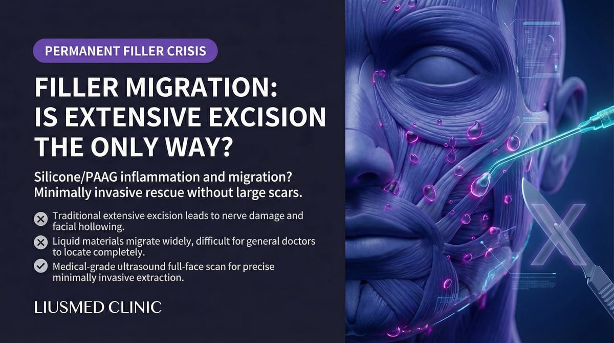
Why Traditional Wide Excision Is Not the Best Approach
The Dilemma of Open Surgery
When faced with permanent filler complications, many surgeons instinctively recommend excision. However, wide surgical excision carries significant drawbacks:
| Issue With Traditional Surgery | Specific Impact |
|---|---|
| Large incisions | Visible facial scarring |
| Tissue sacrifice | Normal tissue removed along with filler |
| Facial depressions | Severe volume deficit possible after excision |
| Nerve damage risk | Wide dissection may injure facial nerves |
| Prolonged recovery | Weeks to months of swelling and healing |
| Incomplete removal | Material dispersed through tissue may still remain |
Why Doing Nothing Is Also Problematic
Some patients choose to coexist with their permanent fillers, but this carries ongoing risks:
- Continuous immune system stimulation by the foreign material
- Cumulative infection risk over time
- Ongoing filler migration
- Tissue changes that may interfere with future medical imaging
- Psychological burden of persistent concern about filler status
Ultrasound-Guided Minimally Invasive Extraction: The Precise Middle Path
Ultrasound Identification of Permanent Fillers
Different permanent fillers display distinct characteristics on ultrasound imaging:
| Filler Type | Ultrasound Appearance | Identification Difficulty |
|---|---|---|
| Liquid silicone | Scattered hyperechoic dots in a "snowstorm" pattern | Moderate (requires differentiation from normal tissue) |
| PAAG | Irregular hypoechoic areas, possibly septated | Relatively easy (gel contrasts well with tissue) |
| Paraffin | Irregular echogenic masses, possible calcification | Relatively easy |
| Artificial bone powder | Hyperechoic granular deposits | Easy |
Advantages of Minimally Invasive Extraction
Compared to traditional wide excision, ultrasound-guided minimally invasive extraction offers clear advantages:
| Factor | Traditional Excision | Ultrasound-Guided Extraction |
|---|---|---|
| Incision size | 3–10cm | 1–3mm pinhole |
| Normal tissue preservation | Poor | Maximized |
| Scarring | Visible | Nearly invisible |
| Recovery time | 2–6 weeks | 3–7 days |
| Nerve damage risk | Higher | Significantly reduced |
| Staged treatment | Poorly suited | Well suited (minimal burden per session) |
Staged Extraction Strategy
For permanent fillers — especially widely dispersed liquid silicone — staged extraction is often the safer approach:
First Session
- Complete ultrasound assessment and mapping
- Remove filler from primary concentration areas
- Evaluate tissue response
4–8 Week Interval
- Allow tissue recovery and remodeling
- Repeat ultrasound to assess remaining deposits
Subsequent Sessions
- Target residual deposits with refined extraction
- Continue ultrasound monitoring until clinical goals are met
Key Insight: Managing permanent fillers does not require achieving perfect removal in a single session. A staged, precise, minimally invasive approach maximizes filler removal while minimizing tissue trauma.
Site-Specific Considerations
Face (Nose, Chin, Forehead, Temples)
The face is the most common injection site for permanent fillers. Extraction requires particular attention to:
- Facial nerve anatomy: Ultrasound identifies and avoids nerve pathways
- Vascular structures: Facial artery, supraorbital artery, and other vessels must be protected
- Skin thickness: Some areas (such as the nasal dorsum) have thin skin requiring careful technique
- Cosmetic placement: Pinhole entries are placed in concealed locations
Post-Procedure Care
- Ice application for 48 hours to reduce swelling
- Avoid vigorous exercise for one week
- Attend scheduled follow-up ultrasound appointments
- Return immediately for any increasing redness, pain, or fever
When to Seek Evaluation
If any of the following apply to you, professional evaluation is recommended as soon as possible:
- You received cosmetic injections years ago with unknown materials
- The injection site shows recurrent redness, swelling, or inflammation
- You can feel hard lumps or sense that filler has shifted position
- The skin over the injection site has changed color or texture
- You are concerned about the long-term safety of permanent filler in your body
The first step is a comprehensive ultrasound evaluation to determine the filler type, distribution, and current condition.
Further reading:
- Why Fillers Migrate
- Minimally Invasive Filler Lump Extraction Technique
- Why Dissolving Enzymes Fail When Capsules Form
Common questions
Why does cosmetic injection from decades ago only cause problems now?
Because these materials don't fade with time — quite the opposite. Things like liquid silicone, PAAG, and paraffin can't be absorbed once injected. As you age and tissue loosens and your immune state shifts, the recurrent inflammation, migration, and granulomas they trigger tend to get more pronounced. Silicone's delayed granulomas alone can surface anywhere from 5 to 30 years after the injection.
Why are liquid silicone and PAAG especially hard to deal with?
Because, unlike hyaluronic acid, they have no clean boundary. Liquid silicone seeps into the gaps between surrounding tissue and mixes in with normal tissue, and it slowly migrates downward with gravity, so it's hard to lift out "as one mass." PAAG is a gel that spreads along tissue planes and can become a breeding ground for bacteria, and its degradation product (acrylamide) is classified as a possible carcinogen. Neither can be dissolved with an enzyme.
Does treating these always mean wide excision?
Not necessarily — and wide excision usually isn't the better option. A large incision leaves a visible facial scar, can take normal tissue out along with the filler and cause hollowing, and may even injure a nerve. Ultrasound-guided minimally invasive removal is the middle path — going in and out through a 1–3 mm entry, using ultrasound to locate the material and steer around nerves and vessels while preserving as much normal tissue as possible.
Can it all be removed in one go?
There's no need to force a complete removal in a single session. For wider spread — liquid silicone especially — staged removal is often safer: the first session addresses the main concentrated area, then a 4–8 week gap lets tissue recover and remodel, and ultrasound reassesses what's left before finely removing the rest. The aim is to remove the most while keeping tissue damage to a minimum, not to push it all at once.
Is it okay to just leave it alone?
You can choose to coexist with it, but it helps to know the ongoing risks: the immune system stays chronically irritated, infection risk accumulates over time, the filler may keep migrating, and it can also interfere with reading future medical imaging. Whether to treat depends on how much your symptoms and concerns weigh on you. A good first step is an ultrasound evaluation to see clearly what the material is, how it's distributed, and its current state, before deciding on the next move.
About the Author
Dr. Ta-Ju Liu
- Current Position: Director, Liusmed Clinic
- Specialties: Minimally invasive surgery, filler complication repair, ultrasound-guided extraction
- Experience: 15+ years of clinical minimally invasive surgery; over 10,000 successful cases
- Philosophy: "The legacy of early cosmetic injections leaves many patients living with anxiety. They need to know that wide excision is not the only path. Ultrasound-guided minimally invasive extraction allows us to gradually remove materials that should not remain in the body — while protecting facial tissue every step of the way."

Specialties
Credentials
- Kaohsiung Medical University, School of Medicine
- Attending Physician, Dermatology, Kaohsiung Chang Gung Memorial Hospital
- Attending Physician, Aesthetic Center, Kaohsiung Chang Gung Memorial Hospital
- Visiting Physician, Dermatology, Xiamen Chang Gung Hospital
- Visiting Physician, Aesthetic Center, Xiamen Chang Gung Hospital
"For every surgery, I strive to achieve a good outcome through a small incision and refined technique. Minimally invasive surgery is not just a technique — it's a commitment of respect to every patient."
Recovery after any procedure needs peer support too
Want to learn more?
Schedule a consultation for professional evaluation and advice