Filler Lump Extraction: Precision Removal While Preserving Normal Tissue
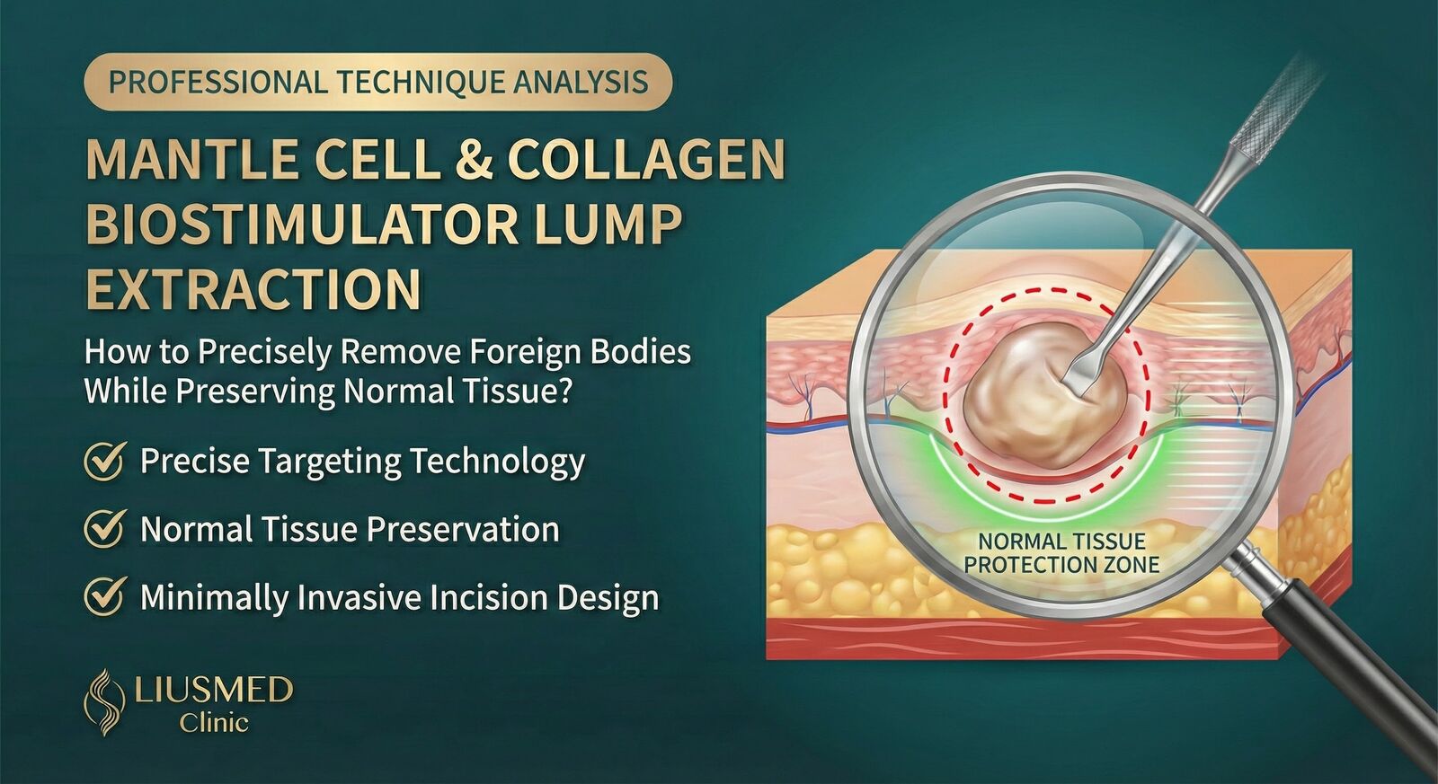
The Core Challenge of Filler Lump Extraction
The goal of minimally invasive filler lump extraction is to completely remove the foreign material while maximally preserving normal tissue. This requires precise localization, skilled technique, and deep understanding of different filler characteristics. This article explains professional extraction techniques and principles.
Types of Filler Lumps
Classification by Filler Material
| Filler Type | Dissolvable | Lump Characteristics | Removal Difficulty |
|---|---|---|---|
| Hyaluronic Acid | ✅ Yes | Soft, clear boundaries | Lower |
| Collagen | ❌ No | Medium firmness | Medium |
| Radiesse (CaHA) | ❌ No | White, granular | Medium-High |
| Collagen Stimulator | ❌ No | Hard, irregular | High |
| Silicone/Oil | ❌ No | Migrating, diffuse | Very High |
| PMMA | ❌ No | Permanent, encapsulated | Very High |
Classification by Problem Type
| Problem Type | Common Cause | Treatment Approach |
|---|---|---|
| Migrated lump | Post-injection displacement | Locate and extract |
| Overfilled lump | Excessive injection | Partial or complete removal |
| Inflammatory lump | Foreign body reaction | Anti-inflammatory then extract |
| Infected lump | Bacterial infection | Control infection first |
| Calcified lump | Long-standing presence | If minimally invasive extraction is not feasible after evaluation, surgical excision may be used |
Why Is Lump Extraction Difficult?
Technical Challenges
| Challenge | Explanation |
|---|---|
| Localization difficulty | Filler may migrate and diffuse, hard to determine extent |
| Unclear boundaries | Mixed with normal tissue, difficult to separate |
| Sensitive location | Face has important nerves and vessels |
| Secondary damage | Over-extraction causes depression |
| Residue risk | Incomplete removal leads to recurrence |
Difficulty Varies by Material
💡 Dr. Liu explains: "Every filler type requires a different extraction approach. Hyaluronic acid can be dissolved first, Radiesse needs curettage, collagen stimulator requires careful separation of fibrotic tissue. No single method works for all cases."
Ultrasound-Guided Technology
Why Is Ultrasound Necessary?
| Advantage | Explanation |
|---|---|
| Precise localization | Real-time imaging shows filler location |
| Boundary identification | Distinguishes foreign material from normal tissue |
| Depth assessment | Understands filler depth and extent |
| Safe guidance | Avoids important vessels and nerves |
| Intraoperative monitoring | Confirms complete removal |
Ultrasound Characteristics of Different Fillers
| Filler | Ultrasound Features |
|---|---|
| Hyaluronic Acid | Hypoechoic, homogeneous |
| Radiesse | Hyperechoic, granular |
| Collagen Stimulator Lump | Mixed echogenicity, irregular |
| Silicone | Hyperechoic, "snowflake" phenomenon |
| Calcified Tissue | Strong echo, posterior shadow |
Precision Extraction Technique
Surgical Principles
- Minimal incision: Use minimally invasive incisions to reduce scarring
- Precise localization: Confirm position under ultrasound guidance
- Conservative excision: Remove only foreign material, preserve normal tissue
- Layer separation: Separate along tissue planes
- Confirm completeness: Ultrasound verification of no residue
Surgical Steps
Step 1: Pre-operative Assessment
| Item | Content |
|---|---|
| History taking | Filler type, injection time, clinic |
| Physical exam | Lump location, size, depth |
| Ultrasound exam | Precise localization and extent assessment |
| Treatment plan | Determine optimal extraction method |
Step 2: Anesthesia and Preparation
- Local anesthesia plus gentle pain-relief anesthesia
- Mark incision location
- Ultrasound preparation
Step 3: Pinhole Design
We use pinhole minimally invasive extraction, with pinhole size typically less than 1.5mm.
| Consideration | Design Principle |
|---|---|
| Hidden scar | Along skin lines or concealed areas |
| Close to lesion | Shorten operating distance |
| Adequate view | Ensure complete extraction possible |
| Minimal wound | Usually < 1.5mm |
Step 4: Lump Extraction
Step 5: Post-operative Care
- Pressure dressing
- Antibiotics for infection prevention
- Schedule follow-up
Extraction Strategies by Filler Type
Hyaluronic Acid Lumps
| Approach | Suitable Situation |
|---|---|
| Hyaluronidase injection | Mild irregularity, small area |
| Dissolve + curettage | Incomplete dissolution residue |
| Direct extraction | Encapsulated, hyaluronidase ineffective |
About Hyaluronidase:
- Can dissolve hyaluronic acid, but limited effect on encapsulated lumps
- Takes effect 24-48 hours after injection
- If ineffective, surgical extraction needed
Radiesse Lumps
| Characteristic | Approach |
|---|---|
| Granular | Primarily curettage |
| May spread | Need careful clearing of surroundings |
| Not dissolvable | Physical removal only |
Collagen Stimulator Lumps
| Characteristic | Difficulty |
|---|---|
| Stimulates tissue growth | Lump and normal tissue hard to distinguish |
| Severe fibrosis | Unclear boundaries |
| May continue growing | Requires complete removal |
Key Points:
- Complete excision of fibrotic tissue
- Ultrasound confirmation of extent
- Post-op monitoring for recurrence
Silicone/Silicone Oil
| Characteristic | Extremely Difficult |
|---|---|
| Migrating diffusion | Cannot completely clear |
| Mixed with tissue | Extraction damages normal tissue |
| Often illegal injection | Very high risk |
⚠️ Warning: Silicone oil filler extraction is extremely difficult, usually can only improve rather than cure, and may require multiple surgeries. The best strategy is "prevention"—don't accept injections from unknown sources.
How to Preserve Normal Tissue?
Technical Keys
| Technique | Explanation |
|---|---|
| Layer recognition | Understand anatomical layers, separate correctly |
| Blunt dissection | Use blunt instruments when possible, reduce bleeding |
| Conservative excision | Only remove confirmed foreign material |
| Intraoperative confirmation | Ultrasound real-time check for residue |
| Staged approach | Multiple sessions if needed to avoid excess damage |
Avoiding Over-Extraction
Over-extraction can cause:
- Tissue depression
- Functional impact (lips, nose)
- Unnatural appearance
- Need for secondary filling
💡 Dr. Liu's Philosophy: "I'd rather do two sessions than extract too much at once. Depression from over-extraction is harder to fix than the original lump."
Frequently Asked Questions
Q1: Can hyaluronic acid lumps be dissolved with hyaluronidase?
A1: It depends:
- Newly injected HA (Hyaluronic Acid) (< 1 year): Usually dissolvable
- Old or encapsulated: Hyaluronidase has limited effect, may need surgery
- Mixed with other fillers: Hyaluronidase only works on HA
Recommend trying dissolution first; consider surgery if ineffective.
Q2: Will minimally invasive extraction leave scars?
A2: Minimally invasive extraction pinholes are usually very small (1-2mm) and placed in concealed locations where possible (hairline, inside nostril, inside mouth). They heal in about 2 weeks and are usually inconspicuous afterwards, although scarring still depends on the individual.
Q3: Will my face look sunken after extraction?
A3: With proper extraction, there won't be obvious depression. But if the original lump was large or in a special location, post-extraction may require:
- Waiting for tissue recovery (about 3 months, reassessed at follow-up)
- Modest filling (using safe hyaluronic acid)
Q4: Is general anesthesia needed?
A4: General anesthesia is not needed. Extraction is done under local anesthesia plus gentle pain-relief anesthesia; during the procedure the doctor talks with you and can pause or adjust at any time.
Q5: Will the lump grow back after minimally invasive extraction?
A5:
| Filler Type | Recurrence Risk |
|---|---|
| Hyaluronic Acid | Very low (won't recur if completely removed) |
| Radiesse | Low (Liusmed minimally invasive technique removes it cleanly) |
| Collagen Stimulator | Low (Liusmed minimally invasive technique removes it cleanly) |
| Ellansé | Low (Liusmed minimally invasive technique removes it cleanly) |
| Silicone | High (often cannot completely clear, but can significantly reduce silicone residue) |
Collagen stimulator lumps require particularly close follow-up.
Post-Op Recovery and Follow-Up
Post-Op Timeline
| Period | Status and Care |
|---|---|
| Days 1-3 | Swelling peak, ice to reduce swelling |
| Week 1 | Swelling subsiding, return for evaluation |
| Week 2 | Suture removal (if applicable) |
| Month 1 | Tissue recovering |
| Month 3 | Evaluate final result |
| 6-12 months | Long-term follow-up (especially for collagen stimulator) |
Guidelines
| ✅ OK | ❌ Avoid |
|---|---|
| Ice to reduce swelling | Heat (first 3 days) |
| Normal diet | Irritating foods |
| Light activity | Strenuous exercise (2 weeks) |
| Attend follow-ups | Self-treating problems |
Conclusion
| Key Point | Explanation |
|---|---|
| Core principles | Precise localization, conservative excision, confirm completeness |
| Technical support | Ultrasound guidance greatly improves precision |
| Material differences | Different fillers need different strategies |
| Prevention is best | Choose reputable clinics, legitimate fillers |
Related Reading
- When Can Failed Aesthetics Be Repaired? Ultrasound-Guided Evaluation
- Complete Guide to Filler Repair
- Post-Aesthetic Golden 7 Days
About the Author
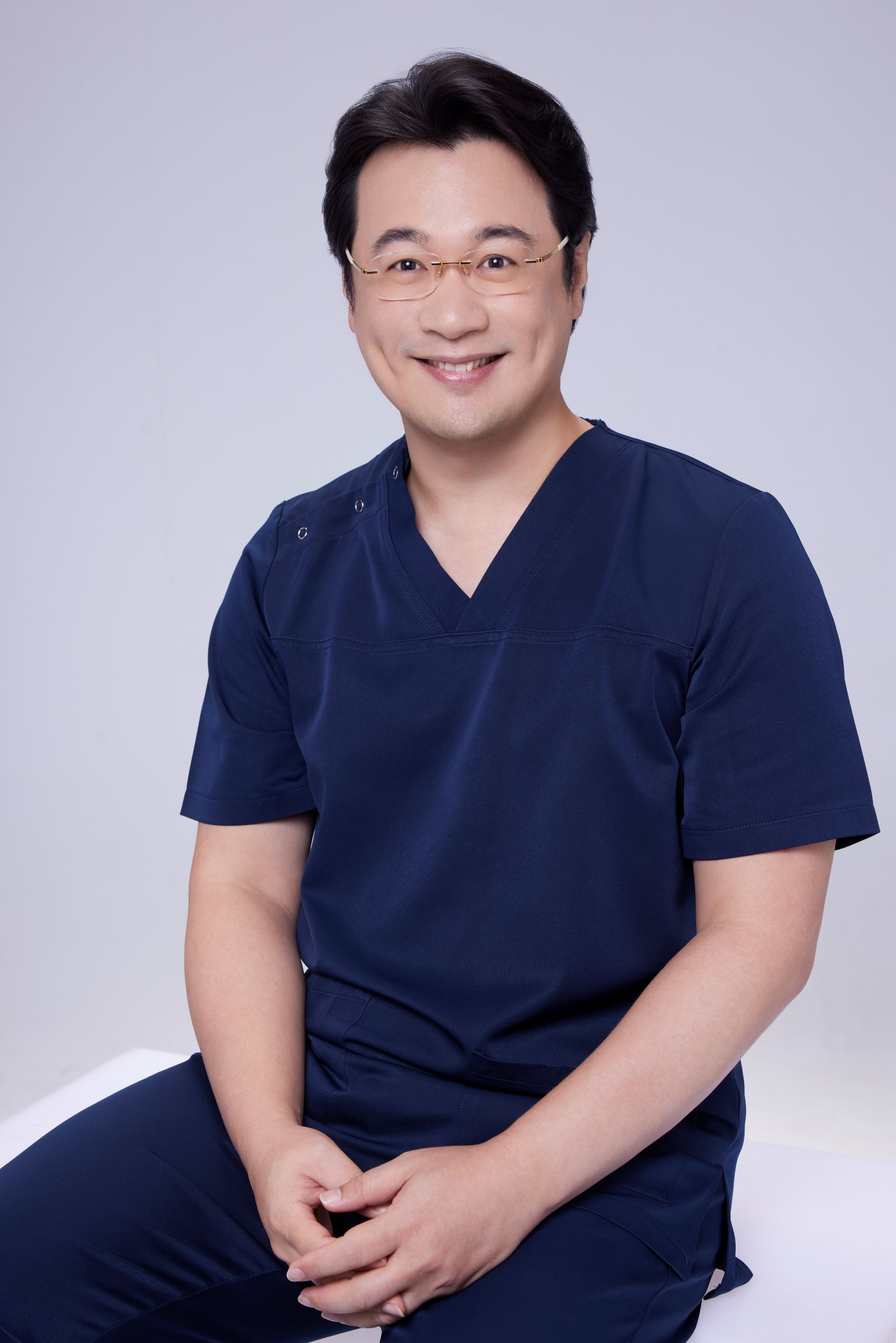
Dr. Ta-Ju Liu
- Current Position: Director, Liushi Clinic
- Specialties: Minimal incision surgery (lipoma, cyst), hyperhidrosis surgery, thread lifting
- Experience:
- 15+ years of clinical minimal incision surgery experience
- Over 10,000 successful minimal incision cases
- Board-certified dermatologist
- Philosophy: "Filler lump treatment requires precision and patience. My goal is to remove the problem while preserving the patient's natural appearance."
Related Services
Specialties
Credentials
- Kaohsiung Medical University, School of Medicine
- Attending Physician, Dermatology, Kaohsiung Chang Gung Memorial Hospital
- Attending Physician, Aesthetic Center, Kaohsiung Chang Gung Memorial Hospital
- Visiting Physician, Dermatology, Xiamen Chang Gung Hospital
- Visiting Physician, Aesthetic Center, Xiamen Chang Gung Hospital
"For every surgery, I strive to achieve a good outcome through a small incision and refined technique. Minimally invasive surgery is not just a technique — it's a commitment of respect to every patient."
Recovery after any procedure needs peer support too


Want to learn more?
Schedule a consultation for professional evaluation and advice