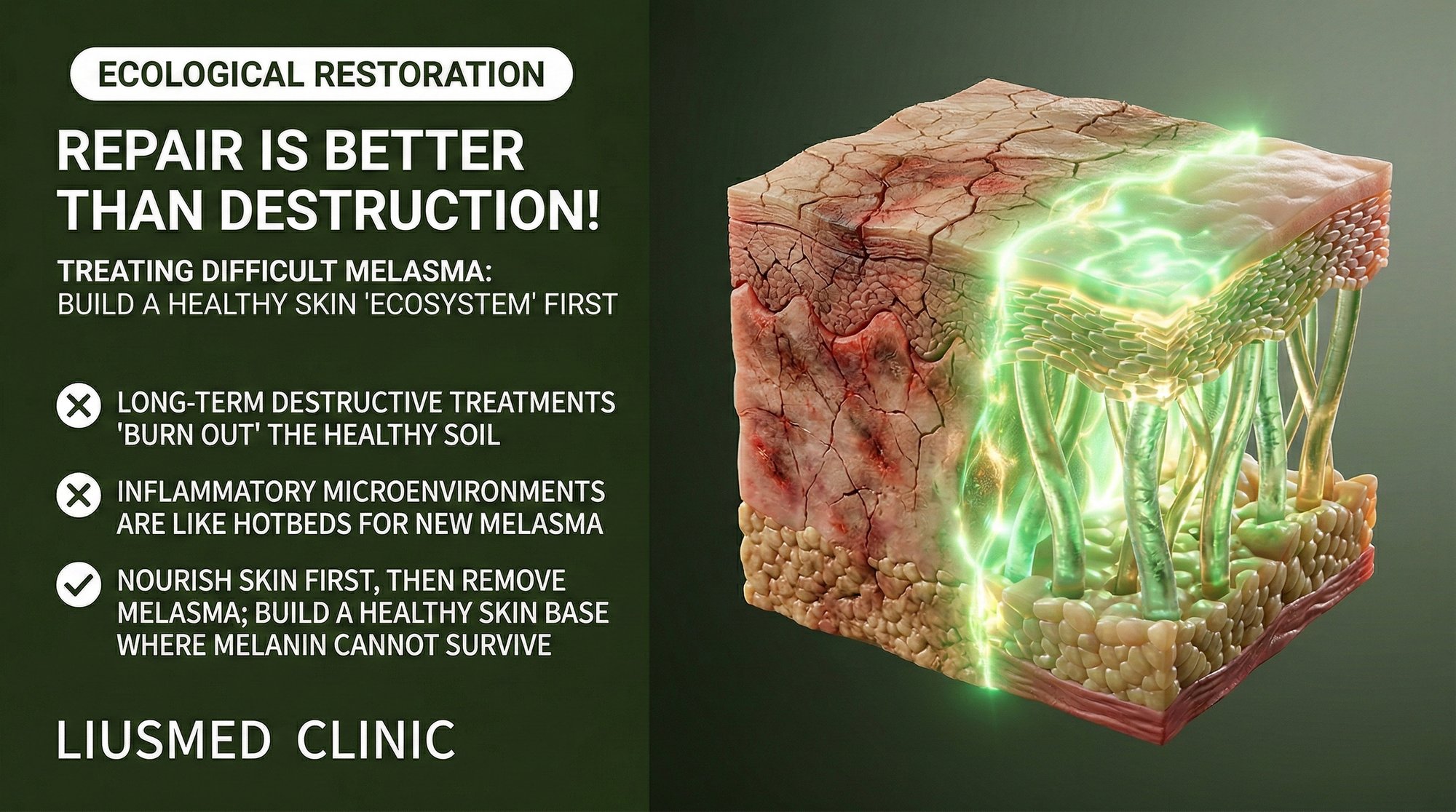
Repair Over Destruction: Why Treating Refractory Melasma Requires Rebuilding the Skin's Healthy Ecosystem
After years of chasing melasma with one destruction-based treatment after another—lasers, peels, aggressive topicals—many patients arrive at a familiar dead end. The patches lighten temporarily, return within months, and sometimes come back worse than before. The frustration is compounded by a nagging question: if these treatments are scientifically proven to destroy melanin and suppress melanocytes, why does the pigmentation keep coming back?
The answer requires a perspective shift. Melasma is not a single-component malfunction. It is a systemic breakdown of the skin's internal ecosystem—a complex, interdependent network of cells, signals, and structures that, when healthy, maintains pigmentation within normal bounds. Destroying one component (melanin) without restoring the ecosystem merely creates a temporary absence of the symptom while the dysfunctional system continues generating the conditions for its return.
This article presents the ecosystem model of melasma and explains why sustainable treatment requires rebuilding rather than destroying.
Table of Contents
- The Skin as an Ecosystem: Key Players
- How the Ecosystem Breaks Down in Melasma
- Why Destruction-Based Approaches Fail Long-Term
- The Repair Framework: Restoring Balance Layer by Layer
- Destruction vs. Repair: A Paradigm Comparison
- What a Repair-First Treatment Journey Looks Like
The Skin as an Ecosystem: Key Players
To understand why melasma is an ecosystem disease, we must first understand what a healthy skin ecosystem looks like. The relevant players include:
Melanocytes Pigment-producing cells that sit at the basal layer of the epidermis. In healthy skin, they produce melanin in response to UV exposure at a rate that protects underlying cells without causing visible hyperpigmentation. They communicate constantly with surrounding keratinocytes and receive signals from the dermis below.
Keratinocytes The majority cell type in the epidermis, keratinocytes receive melanin from melanocytes via melanosome transfer and carry it upward as they differentiate and eventually shed from the skin surface. They also produce paracrine signals (endothelin-1, SCF, alpha-MSH) that modulate melanocyte activity. In an unhealthy ecosystem, stressed keratinocytes chronically overstimulate melanocytes.
Fibroblasts Dermal cells responsible for producing and maintaining the extracellular matrix (collagen, elastin, glycosaminoglycans). Healthy fibroblasts support the structural integrity of the dermis and the basement membrane. Senescent fibroblasts—those that have exited the cell cycle but remain metabolically active—shift from building to destroying, secreting inflammatory cytokines and matrix metalloproteinases (MMPs) that degrade surrounding tissue.
The Basement Membrane (DEJ) The structural interface between epidermis and dermis, composed of collagen IV, laminin, nidogen, and other glycoproteins. When intact, it keeps melanin confined to the epidermis and regulates molecular crosstalk between compartments. When damaged, it allows pigment to drop into the dermis and permits unregulated signaling between inflammatory dermal cells and epidermal melanocytes.
Mast Cells Immune cells in the dermis that release histamine, tryptase, and pro-angiogenic factors when activated. In melasma skin, mast cell density and degranulation activity are elevated, contributing to both inflammation and vascular proliferation.
Dermal Vasculature Blood vessels in the papillary dermis deliver nutrients, immune cells, and signaling molecules. In melasma, the vascular network is abnormally expanded (increased VEGF-driven angiogenesis). Endothelial cells lining these vessels secrete melanocyte-activating factors, creating a vascular-pigmentary feedback loop.
The Extracellular Matrix (ECM) The scaffolding in which all dermal cells reside. Its composition and stiffness influence fibroblast behavior, growth factor availability, and cellular communication. Degraded or fibrotic ECM promotes inflammatory cell phenotypes and impairs normal tissue repair.
In a healthy ecosystem, these components exist in dynamic balance. Melanocytes produce appropriate amounts of pigment; fibroblasts maintain the matrix; the DEJ is intact; mast cells are quiescent; vasculature is normal. Melasma represents a state in which this balance has been lost—and it will not be restored by targeting any single component in isolation.
How the Ecosystem Breaks Down in Melasma
The cascade of ecosystem disruption in melasma typically follows a recognizable sequence, though the specific triggers (UV exposure, hormonal changes, genetic predisposition) vary between patients:
Stage 1: Initial Provocation UV radiation, hormonal shifts, or heat exposure triggers an acute inflammatory response in the dermis. Keratinocytes release pro-inflammatory cytokines downward; mast cells degranulate; fibroblasts activate MMP (Matrix Metalloproteinase) production.
Stage 2: Basement Membrane Degradation Chronic MMP activity erodes collagen IV and laminin at the DEJ. The basement membrane becomes patchy and permeable. Melanin begins to leak into the dermis (pigment incontinence). The loss of DEJ integrity also removes the regulatory filter between epidermal and dermal compartments, allowing inflammatory signals to flow more freely between layers.
Stage 3: Fibroblast Senescence Under sustained inflammatory and oxidative stress, a growing proportion of dermal fibroblasts enter senescence. Instead of repairing the ECM and DEJ, they secrete SASP (Senescence-Associated Secretory Phenotype) factors—IL-6 (Interleukin-1 beta / Interleukin-6), IL-8, MMPs, VEGF (Vascular Endothelial Growth Factor — new blood vessel signal)—that amplify every other pathological process. The very cells that should be rebuilding the tissue are now actively destroying it.
Stage 4: Vascular Remodeling VEGF from senescent fibroblasts and degranulating mast cells drives new blood vessel formation in the papillary dermis. These abnormal vessels deliver more inflammatory cells and signaling molecules to the melasma zone. Endothelial cells secrete endothelin-1 and SCF, directly stimulating melanocytes. The vascular expansion becomes self-sustaining.
Stage 5: Chronic Melanocyte Hyperactivation Receiving persistent stimulation from multiple sources—stressed keratinocytes above, SASP-secreting fibroblasts below, mast cell mediators, and endothelial signals from expanded vasculature—melanocytes settle into a state of chronic overproduction. Even if UV exposure is minimized, the internal signaling environment is sufficient to maintain elevated melanogenesis.
Stage 6: Self-Perpetuating Loop The degraded DEJ fails to contain melanin, allowing continued pigment incontinence. Dermal melanin triggers macrophage recruitment, adding another inflammatory cell population. The inflamed dermis further stimulates fibroblast senescence. The system has reached a self-sustaining pathological equilibrium that persists independently of the original trigger.
Understanding this cascade explains why removing melanin at any single stage does not resolve the disease. The melanin is a downstream product of an upstream ecosystem failure.
Why Destruction-Based Approaches Fail Long-Term
Conventional melasma treatments can be mapped onto the ecosystem model to understand their limitations:
Laser Treatment Targets melanin granules (Stage 5 symptom) while adding thermal energy that can worsen dermal inflammation (Stage 1), accelerate fibroblast senescence (Stage 3), trigger mast cell degranulation (Stage 4), and further damage the DEJ (Stage 2). The short-term pigment clearance is real, but the treatment aggravates multiple upstream nodes of the disease cycle.
Chemical Peels Remove pigmented epidermal cells (Stage 5 symptom) by controlled chemical injury. Superficial peels affect only the epidermis and provide modest, temporary improvement. Deeper peels can breach the DEJ, risking further basement membrane damage (Stage 2) and post-inflammatory melanocyte activation.
Topical Hydroquinone Inhibits tyrosinase in melanocytes (Stage 5 mechanism), slowing melanin production at the cellular level. Does not address dermal inflammation, senescent fibroblasts, vascular changes, or DEJ damage. When discontinued, melanocytes return to baseline overproduction because the signaling environment has not changed. Long-term use carries risks of ochronosis (paradoxical darkening).
Topical Retinoids Accelerate epidermal turnover (shedding pigmented keratinocytes) and may modestly support DEJ component synthesis. However, they do not reach the mid-dermis where the core inflammatory process resides, and their irritant potential can paradoxically trigger inflammatory melanogenesis in sensitive melasma skin.
None of these approaches address the ecosystem as a whole. Each treats one or two nodes while leaving the self-sustaining pathological loop intact.
The Repair Framework: Restoring Balance Layer by Layer
The repair-first approach to refractory melasma—as practiced through the Melasma Injection Treatment at Liusmed Clinic—systematically addresses each layer of the dysfunctional ecosystem:
Layer 1: Quench Dermal Inflammation Intradermal delivery of anti-inflammatory agents (tranexamic acid, specific anti-inflammatory peptides) directly into the upper dermis targets the inflammatory core. Tranexamic acid inhibits plasminogen activation—a key amplifier of the inflammatory cascade—while also reducing mast cell activation and VEGF expression. By delivering these agents via manual injection rather than relying on topical penetration, therapeutic concentrations are achieved precisely where they are needed.
Layer 2: Stabilize and Repair the DEJ With inflammation reduced and MMP activity suppressed, the basement membrane can begin to regenerate. Growth factors and signaling peptides delivered intradermally stimulate fibroblast and basal keratinocyte production of collagen IV, laminin-332, and nidogen. A repairing DEJ progressively seals the epidermal-dermal interface, reducing pigment incontinence. For more on this critical structure, see our article on basement membrane damage and melasma recurrence.
Layer 3: Modulate Fibroblast Behavior The goal is to shift the fibroblast population from a senescent, SASP-secreting phenotype back toward a regenerative, matrix-building phenotype. This is achieved through a combination of anti-inflammatory signaling (reducing the stress that drives senescence), growth factor support (promoting healthy fibroblast function), and matrix repair (providing a healthier ECM environment that favors regenerative cellular behavior).
Layer 4: Normalize Vasculature Anti-angiogenic effects of intradermal tranexamic acid, combined with reduced VEGF output from modulated fibroblasts and mast cells, gradually normalize the expanded vascular network. As abnormal vessels regress, the delivery of inflammatory mediators to the melasma zone decreases, further dampening the pigmentary drive.
Layer 5: Allow Melanocyte Recalibration With the upstream signals quieted—inflammation reduced, mast cells stabilized, SASP suppressed, vessels normalized—melanocytes gradually recalibrate their output toward normal levels. This is not a forced suppression (as with hydroquinone) but a natural recalibration in response to a normalized signaling environment. The result is more physiologic and more sustainable.
Layer 6: Maintain and Protect Rigorous photoprotection (broad-spectrum UV plus visible light), barrier support, and periodic maintenance treatments preserve the restored ecosystem. The goal is to prevent the cascade from re-initiating, not to perpetually suppress its end product.
Destruction vs. Repair: A Paradigm Comparison
| Dimension | Destruction Paradigm | Repair Paradigm |
|---|---|---|
| Disease Model | Melasma = excess melanin | Melasma = ecosystem dysfunction |
| Primary Target | Melanin granules, melanocyte enzyme activity | Dermal inflammation, DEJ integrity, fibroblast health, vasculature |
| Therapeutic Mechanism | Fragment, bleach, exfoliate | Anti-inflammatory delivery, matrix repair, signal normalization |
| Thermal/Chemical Load | Adds energy or chemical stress to tissue | Zero thermal load; delivers reparative agents |
| Effect on Disease Cycle | Interrupts at downstream symptom; may aggravate upstream nodes | Addresses upstream causes; downstream symptoms resolve naturally |
| Recurrence Pattern | High—pathological loop remains intact | Lower—loop is disrupted at multiple nodes |
| Melanocyte Response | Forced suppression or destruction | Natural recalibration via normalized environment |
| DEJ Impact | Risk of further damage | Active repair and restoration |
| Long-Term Tissue Health | Potential cumulative damage (guttate hypomelanosis, thinning) | Improved tissue quality, healthier ECM |
| Patient Experience | Fast initial results, frustrating recurrence cycles | Gradual improvement, progressive stability |
What a Repair-First Treatment Journey Looks Like
For patients considering the repair-first approach, understanding the treatment arc helps set appropriate expectations:
Weeks 1-4: Foundation Phase Initial assessment including melasma mapping (distribution, depth estimation, inflammatory activity). First two to three injection sessions focus on delivering anti-inflammatory and anti-angiogenic agents into the active dermis. Visible changes are subtle at this stage—reduced redness and skin "calming" are often the first signs, reflecting decreased dermal inflammation rather than pigment clearance.
Weeks 4-12: Active Repair Phase Continued injection sessions at two to four week intervals. DEJ repair begins, pigment incontinence slows, and dermal melanin undergoes gradual clearance as the inflammatory drive diminishes. Patients typically notice progressive lightening that builds incrementally rather than the dramatic "before and after one session" pattern seen with laser treatments. The improvement, however, is more stable.
Months 3-6: Consolidation Phase Treatment frequency decreases as the dermal environment stabilizes. Melanocytes recalibrate toward normal output. Residual dermal melanin continues to clear slowly via melanophage turnover. The comparison between the patient's current state and their pre-treatment baseline becomes increasingly apparent.
Ongoing: Maintenance Phase Periodic maintenance sessions (every two to four months, depending on individual response) combined with rigorous photoprotection and topical barrier support. The goal is to sustain the restored ecosystem and prevent recurrence triggers (UV, heat, hormonal fluctuations) from re-initiating the inflammatory cascade.
This timeline is longer than a single laser session, but the outcome is fundamentally different: instead of temporary pigment removal in a still-dysfunctional tissue, the result is a genuinely healthier dermal environment in which melasma has less opportunity to recur.
Frequently Asked Questions
Q1: If melasma is an ecosystem problem, why do some people get better with just sunscreen and hydroquinone?
Patients with mild, predominantly epidermal melasma and minimal dermal involvement may respond to surface-level interventions because their ecosystem disruption is relatively contained. The deeper the dermal involvement—the more senescent fibroblasts, vascular changes, DEJ damage, and mast cell activity—the less likely that topical-only approaches will provide lasting benefit. The ecosystem model is most relevant for refractory melasma that has failed conventional treatments.
Q2: How do I know if my melasma is "ecosystem-level" dysfunction versus simple surface pigmentation?
Key indicators include: melasma duration exceeding two years, resistance to at least two previous treatment modalities, visible rebound within three to six months of treatment, a bluish-gray (rather than brown) hue suggesting dermal pigment, and worsening after laser treatments. A clinical evaluation at a specialized practice like Liusmed Clinic can assess these factors and determine the appropriate treatment tier.
Q3: Can the ecosystem model explain why my melasma worsened after pregnancy even though it was previously stable?
Yes. Pregnancy introduces hormonal shifts (elevated estrogen and progesterone) that can re-trigger the inflammatory cascade at Stage 1, even in skin that had reached a stable state. If the DEJ was already mildly compromised (subclinical damage), the hormonal provocation can push the ecosystem past a tipping point—activating mast cells, stimulating vascular changes, and driving melanocytes into overproduction. This is why post-partum melasma can be more severe than the pre-pregnancy baseline.
Q4: Is the Melasma Injection Treatment suitable for all skin types?
The repair-first approach is suitable for all Fitzpatrick (Fitzpatrick Skin Type) skin types (I-VI). In fact, it may be particularly advantageous for darker skin types (IV-VI) where laser treatments carry elevated risks of post-inflammatory hyperpigmentation and hypopigmentation. Because the injection protocol does not involve thermal energy or surface-level chemical injury, the risks specific to melanin-rich skin are substantially reduced.
Q5: What happens if I stop maintenance treatments after completing the initial repair phase?
The stability of results after completing the repair phase depends on individual factors including ongoing UV exposure, hormonal status, skin care compliance, and the degree of ecosystem recovery achieved. Some patients maintain excellent results with photoprotection alone for extended periods. Others—particularly those with strong hormonal triggers or occupational sun exposure—benefit from periodic maintenance sessions to reinforce the dermal environment. The repair-first approach aims to reduce treatment dependency, not eliminate it entirely.
Q6: Can I combine the repair approach with topical treatments?
Absolutely. Topical agents that support the repair framework—such as barrier-repair moisturizers, gentle retinoids (introduced cautiously), azelaic acid, and vitamin C—can complement the intradermal injection protocol. The key is avoiding topical agents that provoke inflammation (over-exfoliation, high-concentration acids, irritant actives) and maintaining consistent broad-spectrum photoprotection. Your physician can recommend a tailored topical regimen that aligns with the ecosystem repair strategy.
About the Author
Dr. Ta-Ju Liu is the founder of Liusmed Clinic in Taipei, Taiwan, where he leads a practice dedicated to regenerative medicine and minimal incision surgery. With dual expertise in dermatology and surgical repair, Dr. Liu developed the clinic's signature repair-over-destruction philosophy after observing that many refractory skin conditions—including melasma, rosacea, and filler complications—share a common root in chronic tissue-level inflammation. His clinical approach prioritizes restoring the skin's native repair mechanisms rather than adding further injury through aggressive interventional techniques.
Disclaimer
This article is provided for educational and informational purposes only and does not constitute medical advice, diagnosis, or treatment. Individual results vary based on skin type, condition severity, and treatment compliance. Always consult a qualified dermatologist or medical professional before beginning any treatment for melasma or other skin conditions. The information presented reflects the clinical perspective of the author and Liusmed Clinic as of the publication date.

Specialties
Credentials
- Kaohsiung Medical University, School of Medicine
- Attending Physician, Dermatology, Kaohsiung Chang Gung Memorial Hospital
- Attending Physician, Aesthetic Center, Kaohsiung Chang Gung Memorial Hospital
- Visiting Physician, Dermatology, Xiamen Chang Gung Hospital
- Visiting Physician, Aesthetic Center, Xiamen Chang Gung Hospital
"For every surgery, I strive to achieve a good outcome through a small incision and refined technique. Minimally invasive surgery is not just a technique — it's a commitment of respect to every patient."
Recovery after any procedure needs peer support too
Want to learn more?
Schedule a consultation for professional evaluation and advice