AestheFill Lumps and Uneven Texture Six Months After Injection: Treatment and Minimally Invasive Solutions
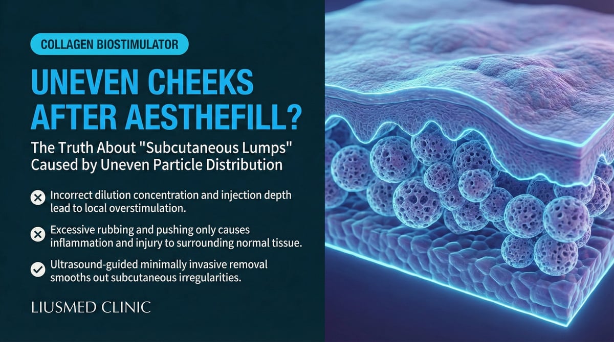
Why Do Cheeks Become Bumpy Months After AestheFill?
AestheFill is a collagen-stimulating filler that has rapidly gained popularity across Asian markets. Its active ingredient — poly-D,L-lactic acid (PDLLA) microspheres — works similarly to Sculptra (PLLA) by stimulating the body's own collagen production to create volumization and skin tightening effects. For the ingredient and brand background of AestheFill as a material, the filler-revision site has an AestheFill (PDLLA) encyclopedia page.
However, a significant number of patients find that three to six months after injection, the anticipated "natural plumpness" transforms into uneven cheek contours, palpable hard lumps, or even visible subcutaneous nodules. The shift from anticipation to distress leaves many patients feeling helpless.
Key Insight: Although AestheFill (PDLLA) and Sculptra (PLLA) share similar chemistry, their microsphere morphology and degradation kinetics differ — and these differences can influence the pattern and severity of complications.
Understanding AestheFill: Composition, Mechanism, and Risks
PDLLA Microsphere Characteristics
The core component of AestheFill is PDLLA microspheres. Unlike the pure L-form PLLA (Poly-L-Lactic Acid (Sculptra) — particle injection stimulating collagen) in Sculptra, PDLLA is a copolymer of both D- and L-lactic acid:
| Property | AestheFill (PDLLA) | Sculptra (PLLA) |
|---|---|---|
| Chemistry | Poly-D,L-lactic acid | Poly-L-lactic acid |
| Crystallinity | Amorphous | Semi-crystalline |
| Degradation rate | Theoretically faster | Theoretically slower |
| Microsphere shape | Porous spheres | Irregular fragments |
| Collagen stimulation | Porous structure promotes cell ingrowth | Surface stimulates collagen formation |
Why Uneven Texture Develops
The causes of post-AestheFill texture irregularity are multifactorial:
-
Technical Factors
- Insufficient dilution: High microsphere concentration promotes aggregation
- Uneven injection depth: Variable placement creates non-uniform collagen stimulation
- Inconsistent injection speed: Some areas receive disproportionately dense deposits
-
Biological Factors
- Individual collagen response varies significantly between patients
- Local blood circulation affects degradation rate
- Tissue tension differences lead to variable collagen growth patterns
-
Microsphere-Related Factors
- Porous structure may provoke localized intense fibrosis
- Microsphere aggregation forms "cores" that become excessively wrapped in collagen
- Non-uniform degradation creates areas of persistent stimulation
Key Insight: Texture irregularity typically becomes apparent 2–6 months post-injection, coinciding with peak collagen production. The window between "looking great" and "growing concerned" can be alarmingly short.
Conventional Treatments and Their Limitations
5-FU (5-Fluorouracil) Injection
5-FU is an antimetabolite medication sometimes used to treat collagen stimulator nodules. It works by inhibiting fibroblast proliferation and reducing excessive collagen formation.
| Aspect | Details |
|---|---|
| Mechanism | Inhibits fibroblast DNA synthesis |
| Best suited for | Early inflammatory nodules |
| Frequency | Every 2–4 weeks |
| Typical course | 3–5 sessions |
| Limitation | Limited effect on mature fibrous capsules |
Steroid Injection
Intralesional steroid injection (triamcinolone) is also commonly attempted to soften nodules:
| Potential Benefit | Risk |
|---|---|
| Anti-inflammatory effect | Skin atrophy |
| Temporary softening | Hypopigmentation |
| Pain reduction | Telangiectasia |
| — | Fat atrophy (especially noticeable on the face) |
When 5-FU and Steroids Fail
Clinical experience shows that 5-FU and steroids have limited effectiveness in the following scenarios:
- Mature encapsulated lumps: Drugs cannot penetrate the fibrous barrier
- Heavily aggregated microsphere nodules: Medication reduces surrounding inflammation but the core material remains
- Multiple deep nodules: Superficial drug injections cannot reach deep lesions
- Nodules older than 6 months: Fibrosis (excess scar tissue formation) has stabilized; drug efficacy diminishes
Ultrasound-Guided Extraction: When Medical Treatment Reaches Its Limit
AestheFill Nodules on Ultrasound
AestheFill nodules display characteristic features on ultrasound imaging:
| Ultrasound Feature | Clinical Significance |
|---|---|
| Well-defined hypoechoic nodule | Indicates capsule formation |
| Hyperechoic dots within the nodule | Residual PDLLA microspheres |
| Altered echogenicity of surrounding tissue | Reflects local fibrosis degree |
| Distance between nodule and skin surface | Determines whether surface irregularity is visible |
Indications for Extraction
Ultrasound-guided minimally invasive extraction should be considered when:
- Three or more 5-FU sessions have produced no significant improvement
- Steroid injections have caused skin atrophy while the lump persists
- Nodules have been present for over 6 months and affect appearance
- The patient cannot accept waiting 2–3 years for natural degradation
- Superficial nodules are severely disrupting facial contour
The Extraction Process
Assessment Phase
- Detailed history: AestheFill batch, injection date, treatment areas, and dosage
- Ultrasound scan: Document all nodule locations, dimensions, depth, and capsule characteristics
- Discuss realistic expectations with the patient
Surgical Phase
- Local anesthesia plus gentle pain-relief anesthesia
- 1–2mm pinhole incision
- Real-time ultrasound-guided approach to the nodule
- Fragment the capsule and extract aggregated PDLLA microspheres
- Ultrasound confirmation of extraction completeness
Post-Procedure Phase
- Light compression for 24–48 hours
- One-week follow-up
- Ultrasound monitoring at 1 month and 3 months
Expected Results
| Nodule Type | Expectation (discussed individually after ultrasound assessment) |
|---|---|
| Single superficial nodule | Usually clear improvement; 100% removal not guaranteed |
| Multiple superficial nodules | Can treat several simultaneously |
| Deep nodules | Good improvement; may require pathway confirmation |
| Diffuse texture irregularity | Noticeable improvement; may need staged treatment |
Preventing AestheFill Complications
Pre-Injection Evaluation
- Understand your skin thickness and tissue conditions
- Confirm the practitioner's experience with AestheFill specifically
- Discuss injection volume (less is more)
- Establish a plan for managing complications if they arise
The Importance of Injection Technique
| Best Practice | Risk Practice |
|---|---|
| Adequate dilution | Insufficient dilution |
| Deep, uniform injection | Variable depth placement |
| Small volumes over multiple sessions | Large single-session volumes |
| Post-injection massage for even distribution | Neglecting post-injection massage |
| Avoiding thin-skinned periorbital area | Injecting in high-risk zones |
Common questions
How soon after AestheFill do lumps or uneven texture usually show up?
Most of the time the unevenness creeps in somewhere between two and six months after the injection, which is right when collagen production peaks. That's why it can look great at first and then start to feel bumpy or show visible nodules a while later. The stretch from happy to worried is often pretty short.
If 5-FU and steroid injections haven't helped, are there other options?
Yes. 5-FU and steroids have a better shot at early inflammatory nodules, but once a lump has formed a mature fibrous capsule, the drugs struggle to get through it and the core material stays put. That's the point where ultrasound-guided extraction becomes a way to deal with the leftover material directly.
How is the ultrasound-guided extraction actually done, and is the wound big?
It's done under local anesthesia plus gentle pain-relief anesthesia through a pinhole of about one to two millimeters. Using real-time ultrasound, we reach the nodule, break up the capsule, take out the clustered PDLLA microspheres, and then check with ultrasound how much came out. The opening is tiny — usually light compression for a day or two, then a follow-up about a week later.
If I just leave the lump alone, will it go away on its own?
PDLLA does break down over time, so in theory a couple of years might soften it. But if the lump is already affecting how your face looks, or you'd rather not wait that long, you don't have to. The main thing is to avoid repeated drug injections, since going back again and again can add tissue damage — getting a proper evaluation early tends to make more sense.
What can I do to lower the risk of lumps in the first place?
A few things help: adequate dilution, spreading the volume over smaller sessions rather than a lot at once, staying away from the thin skin around the eyes, massaging afterward as advised so it settles evenly, and checking that your injector has real experience with this material. The guiding idea is that less is more.
Do Not Let Texture Irregularity Become a Long-Term Burden
If you are experiencing uneven cheeks, lumps, or nodules after AestheFill injection — and medical treatments have not delivered results — ultrasound-guided minimally invasive extraction may be the answer. Early professional evaluation is important to avoid cumulative tissue damage from repeated drug injections.
Further reading:
- Collagen Stimulator Nodules: What to Do When 5-FU Treatment Fails
- Sculptra Lumps: Options After Steroid Failure
- Minimally Invasive Filler Lump Extraction Technique
About the Author
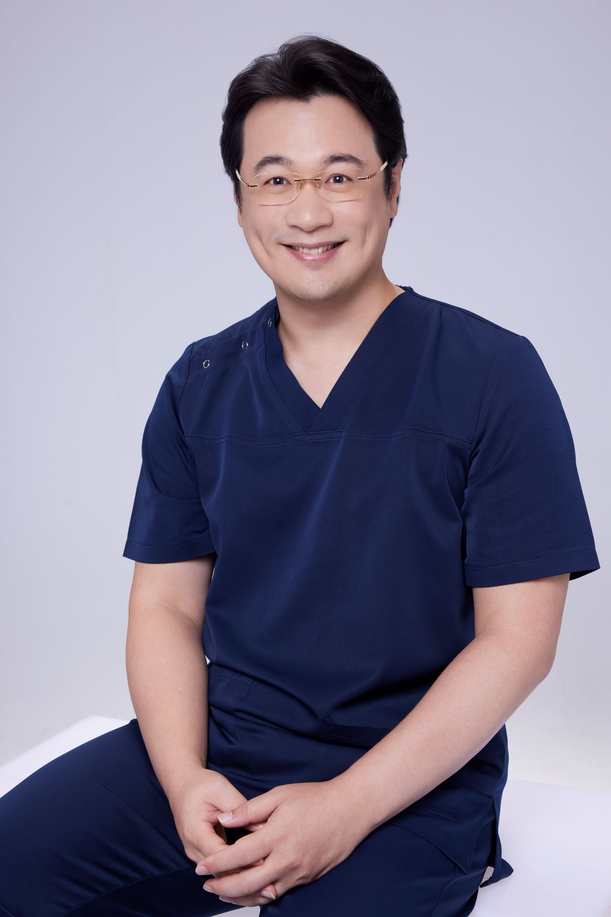
Dr. Ta-Ju Liu
- Current Position: Director, Liusmed Clinic
- Specialties: Minimally invasive surgery, filler complication repair, ultrasound-guided extraction
- Experience: 15+ years of clinical minimally invasive surgery; over 10,000 successful cases
- Philosophy: "Managing collagen stimulator complications requires patience and precision. Medical treatment has its role, but when medication reaches its limit, minimally invasive extraction provides the definitive answer."
Related Services
Specialties
Credentials
- Kaohsiung Medical University, School of Medicine
- Attending Physician, Dermatology, Kaohsiung Chang Gung Memorial Hospital
- Attending Physician, Aesthetic Center, Kaohsiung Chang Gung Memorial Hospital
- Visiting Physician, Dermatology, Xiamen Chang Gung Hospital
- Visiting Physician, Aesthetic Center, Xiamen Chang Gung Hospital
"For every surgery, I strive to achieve a good outcome through a small incision and refined technique. Minimally invasive surgery is not just a technique — it's a commitment of respect to every patient."
Recovery after any procedure needs peer support too


Want to learn more?
Schedule a consultation for professional evaluation and advice