Case Analysis: Severe Ellansé Nodules — Complete Ultrasound-Guided Micro-Extraction Record
Case Scenario
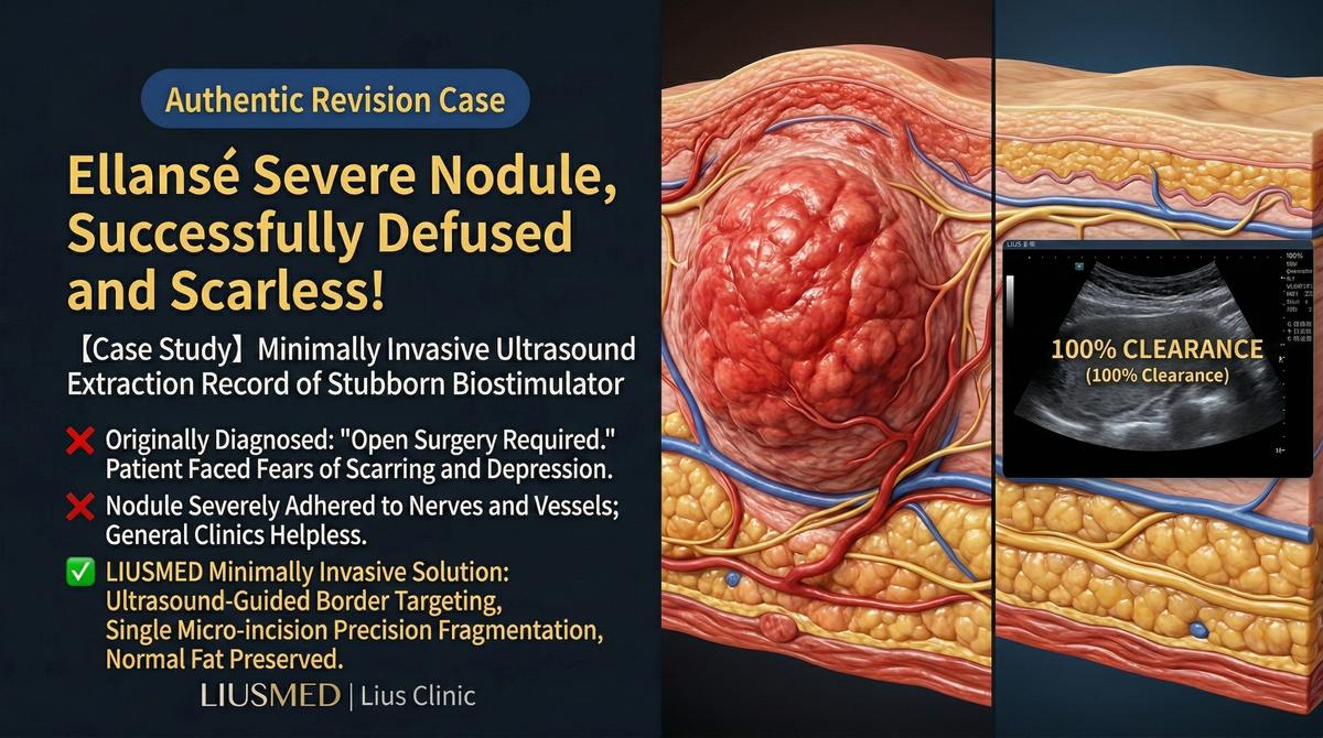
Patient Background: A middle-aged woman who received Ellansé (polycaprolactone) injections in the cheek area approximately two years prior to address cheek hollowing. Approximately 6 months post-injection, palpable lumps developed at the injection sites, gradually enlarging and becoming more visible over time.
Treatment History:
- Returned to the original clinic; told "this is normal collagen stimulation that will resolve on its own"
- After 6 months of waiting with no resolution, sought additional medical attention
- Received 3 sessions of local steroid injections — lumps slightly softened but did not noticeably reduce in size
- Attempted 2 sessions of 5-FU injection with limited effect
- Found information about ultrasound-guided micro-extraction through online research
Presentation at Consultation:
- 1-2 palpable lumps on each cheek
- Slight skin elevation over the lumps, visible at certain angles
- Firm to touch, non-tender
- Approximately one year of conservative treatment with limited results
Deep Analysis
Root Cause Analysis
| Aspect | Finding |
|---|---|
| Material characteristics | Ellansé consists of PCL microspheres suspended in CMC gel; PCL cannot be enzymatically dissolved |
| Nodule etiology | Likely fibrous capsule reaction and excessive collagen stimulation around PCL microspheres |
| Why conservative treatment failed | Steroids can soften fibrous tissue but cannot eliminate PCL microspheres; 5-FU has limited effect on PCL-induced collagen |
| Ultrasound findings | Multiple medium-high echogenicity nodules in subcutaneous fat layer, irregular borders, surrounded by fibrotic bands |
| Vascular assessment | Nodules located at safe distance from major vessels |
Key Insight: Ellansé nodules are fundamentally different from hyaluronic acid nodules — they cannot be "dissolved." Conservative steroid or 5-FU treatment can only soften surrounding tissue but cannot eliminate the core PCL (Polycaprolactone (Ellansé) — longer-lasting collagen stimulator) microspheres. When conservative treatment exceeds one year without significant improvement, physical extraction is a reasonable next step.
Related reading: Can Ellansé Be Removed?
Doctor's Perspective
Dr. Liu's analysis after evaluation:
"This patient's situation represents a typical case among Ellansé complications. Ultrasound clearly revealed multiple nodules, each approximately 8-15mm, wrapped in prominent fibrotic tissue. The positive finding was that the nodules were located in the subcutaneous fat layer at moderate depth, with no major vessels in close proximity — technically feasible for minimally invasive extraction.
Notably, because the nodules had been present for over a year and had undergone multiple steroid injections, the surrounding fibrosis was fairly dense. Greater care would be needed during extraction to separate the filler-tissue boundary. Our goal was to extract the PCL microspheres and their reactive tissue as completely as possible while preserving normal subcutaneous fat."
Treatment Plan and Process
Pre-Operative Planning
| Planning Item | Content |
|---|---|
| Ultrasound marking | Mark all nodule locations, sizes, and depths |
| Vascular mapping | Confirm facial artery and vein pathways |
| Entry point selection | Choose the nearest concealed micro-entry to each nodule |
| Surgical sequence | Address shallower, easier nodules first, then deeper ones |
Surgical Process
- Local anesthesia: Precise local infiltration under ultrasound guidance
- Micro-entry: 1-2mm pinhole as the operative entry
- Ultrasound-guided targeting: Real-time imaging confirms instruments reach the target nodule
- Dissection and extraction: Careful separation of nodule from surrounding fibrotic tissue, progressive removal
- Real-time verification: Immediate ultrasound scan after each nodule extraction
- Residual assessment: Comprehensive scan confirming no significant residual material
Extracted Material Observations
The extracted tissue consisted of firm white to grayish-white masses. Cross-section revealed PCL microsphere particles encased in fibrous tissue, consistent with pre-operative ultrasound findings.
Key Patient Notes
Post-Operative Recovery
| Timeline | Expected Condition |
|---|---|
| Immediately post-op | Mild swelling and bruising at surgical sites |
| Days 1-3 | Peak swelling period, gradually begins to subside |
| Week 1 | Most swelling resolved, bruising begins to fade |
| Week 2 | Noticeable appearance improvement, lumps gone |
| Month 1 | Tissue continues to soften and recover |
| Month 3 | Final results gradually stabilize |
Important Notes
- Post-operative swelling is normal and does not indicate surgical failure
- Bruising may persist 1-2 weeks and can be concealed with makeup
- Temporary tactile changes at the surgical site typically resolve within weeks
- Post-extraction depressions are usually assessed after swelling subsides to determine if further treatment is needed
- Complete recovery requires 2-3 months
Key Insight: Ellansé nodule extraction differs from simple HA (Hyaluronic Acid — sugar molecule naturally in skin, holds water) dissolution — it requires physical manipulation, so the recovery period is longer. However, compared to continuing to live with nodules or pursuing conservative treatments with limited effect, micro-extraction offers a more definitive resolution.
Related reading: When 5-FU Fails for Collagen Stimulator Lumps
Clinical Takeaways from This Case
- Ellansé nodules require timely and accurate assessment — prolonged conservative treatment may worsen fibrosis
- Ultrasound is essential for evaluation and extraction — palpation cannot accurately determine nodule count, size, or depth
- Micro-extraction is a viable option — large open excisions are not necessary
- Patient expectation management is critical — recovery takes time, and final results are not immediate
If you are experiencing similar Ellansé complications, schedule a consultation for an ultrasound evaluation.
Related reading: Filler Lump Extraction Technique
Related Services

Specialties
Credentials
- Kaohsiung Medical University, School of Medicine
- Attending Physician, Dermatology, Kaohsiung Chang Gung Memorial Hospital
- Attending Physician, Aesthetic Center, Kaohsiung Chang Gung Memorial Hospital
- Visiting Physician, Dermatology, Xiamen Chang Gung Hospital
- Visiting Physician, Aesthetic Center, Xiamen Chang Gung Hospital
"For every surgery, I strive to achieve a good outcome through a small incision and refined technique. Minimally invasive surgery is not just a technique — it's a commitment of respect to every patient."
Recovery after any procedure needs peer support too
Want to learn more?
Schedule a consultation for professional evaluation and advice