What Happens to Fillers in Your Tissue After 5 to 10 Years?
What Has Happened to the Filler You Injected?
The hyaluronic acid you had injected into your nasolabial folds five years ago — you assumed your body absorbed it long ago. The collagen stimulator injected into your cheeks eight years ago — you assumed only "your own collagen" remained. The calcium hydroxylapatite injected into your nose ten years ago — you had almost forgotten it existed.
But if you were to undergo a high-resolution ultrasound scan today, you might be surprised to discover that many of those fillers that "should have disappeared" are still present in your tissue — only their form has changed dramatically from the day they were injected.
Key Insight: The fate of fillers inside the body is far more complex than what product labels describe. "Absorbable" does not mean "completely gone," and "duration of 12–18 months" does not mean the filler drops to zero after 18 months. Understanding these long-term changes is the first step toward making informed repair decisions.
The Basic Science of Filler Degradation
Three Pathways of In Vivo Degradation
All fillers injected into tissue face three major degradation pathways:
-
Enzymatic degradation: Specific enzymes in the body — such as hyaluronidase — can break down certain filler materials. This is the primary degradation pathway for hyaluronic acid, but cross-linked structures significantly slow the process.
-
Hydrolysis: Tissue fluid gradually infiltrates the chemical structure of the filler, breaking molecular chains. This is the primary degradation pathway for PCL (Polycaprolactone (Ellansé) — longer-lasting collagen stimulator) (polycaprolactone) and PLLA (Poly-L-Lactic Acid (Sculptra) — particle injection stimulating collagen) (poly-L-lactic acid), typically measured in years.
-
Cell-mediated degradation: Macrophages and foreign body giant cells attempt to engulf and break down filler fragments. This process occurs with all fillers but varies greatly in efficiency depending on material properties.
Factors Influencing Degradation Speed
| Factor | Accelerates Degradation | Slows Degradation |
|---|---|---|
| Cross-linking degree | Low cross-linking | High cross-linking |
| Injection site mobility | High-movement areas (lips, nasolabial folds) | Low-movement areas (temples, forehead) |
| Local blood circulation | Rich blood supply | Limited blood supply |
| Injection volume | Small amounts | Large boluses |
| Immune response intensity | Strong response | Mild response |
| Degree of encapsulation | Not encapsulated | Fully encapsulated |
The 5–10 Year Fate of Different Filler Types
Hyaluronic Acid (HA): The Truth About "Absorbable"
HA (Hyaluronic Acid — sugar molecule naturally in skin, holds water) is marketed as an "absorbable, safe, short-acting" filler, with labeled durations typically between 6 and 18 months. However, clinical and imaging evidence tells a different story.
Years 1–2: Surface Effect Decline
During the first 1–2 years, the filler does degrade — but primarily the non-cross-linked or lightly cross-linked portions. The visible volume effect gradually diminishes, and patients believe "the filler has been absorbed." But the highly cross-linked core structure often persists.
Years 2–5: Residue and Remodeling
Remaining highly cross-linked HA fragments continue to degrade slowly, but the rate has decreased markedly. Meanwhile, these fragments may:
- Become encased in fibrous tissue, forming capsular structures
- Gradually migrate downward due to gravity and muscle activity
- Absorb surrounding water, maintaining a certain volume
- Trigger a low-grade chronic inflammatory response
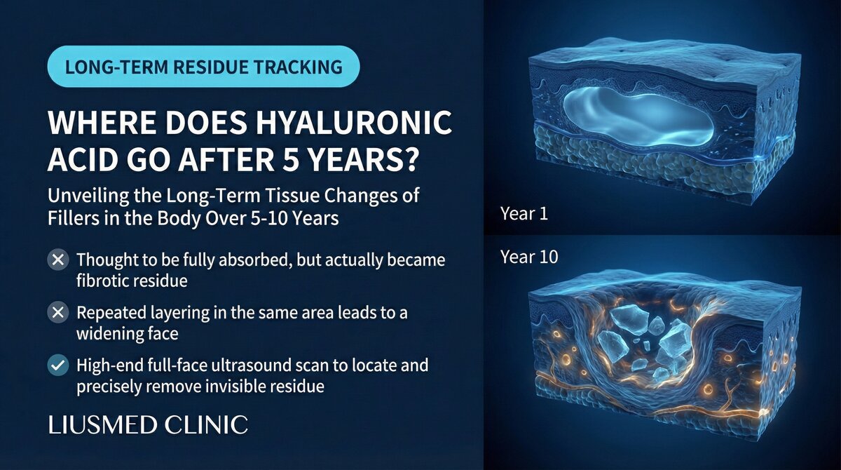
Years 5–10: You Think It Is Gone
On ultrasound, HA injected 5–10 years ago can frequently be detected — as small residual clusters, scattered fragments, or encapsulated nodules. These residues may not cause obvious aesthetic issues, but they are there, and they may interact with new filler if re-injection is performed.
Key Insight: Studies show that even at 2–3 times the labeled duration, a significant proportion of HA residue can be detected on MRI (Magnetic Resonance Imaging) or ultrasound. "Absorbable" does not equal "completely absorbed." To learn more about this myth, see The Myth of Complete HA Absorption.
Poly-L-Lactic Acid (PLLA/Sculptra): The Long-Term Fate of Microspheres
PLLA is injected as microspheres designed to stimulate autologous collagen production. The microspheres themselves are expected to degrade completely within 2 years.
Years 1–3: Microsphere Degradation and Collagen Production in Parallel
PLLA microspheres gradually disintegrate through hydrolysis into lactic acid molecules, which are metabolized by the body. Simultaneously, the microspheres stimulate surrounding collagen neogenesis, producing volume.
Years 3–5: Potential Nodule Formation
If microsphere degradation is uneven (certain areas accumulate too many microspheres), localized nodules of excessive collagen proliferation may form. The cores of these nodules may still contain incompletely degraded PLLA fragments, surrounded by dense collagen fibers.
Years 5–10: Long-Term Persistence of Collagen Structures
Even if PLLA microspheres eventually degrade completely, the collagen structures they stimulated do not disappear along with them. This new collagen may persist for many years, forming permanent fibrotic structures in some cases. Learn more about lumps appearing years after injection.
Polycaprolactone (PCL/Ellanse): The Slow Journey of Microspheres
PCL microspheres degrade more slowly than PLLA, with designed durations of 1–4 years depending on the product variant.
Years 1–3: Designed Action Period
PCL microspheres slowly degrade while stimulating collagen production. The gel carrier (CMC) is absorbed within months, but the microspheres themselves degrade over years.
Years 3–7: Residual Period
Clinical experience shows that some PCL microspheres may persist beyond their designed duration. These residual microspheres may:
- Become encapsulated and remain stable
- Continue degrading at an extremely slow rate
- In certain cases, trigger delayed immune responses
Years 7–10 and Beyond
Long-term follow-up data is limited, but case reports indicate that some PCL residues can be detected more than 7 years after injection. The clinical significance of these residues — whether they will cause problems — depends on individual immune responses and residual volume.
Permanent Fillers: Guests Who Never Leave
Silicone, polyacrylamide gel (PAAG), and other permanent fillers, as their name implies, are never broken down by the body.
| Timeline | Changes | Clinical Impact |
|---|---|---|
| 1–3 years | Stable period, encapsulation may begin | Usually no obvious problems |
| 3–5 years | Capsule gradually thickens, migration may begin | Mild asymmetry, hardened feel |
| 5–10 years | Chronic inflammation, cumulative migration, capsular contracture | Shape distortion, lump formation |
| 10–20 years | Severe migration, granulomas, tissue destruction | Significant deformity, repair surgery needed |
| 20+ years | Deep integration with surrounding tissue | Extremely difficult to remove |
Long-Term Filler–Tissue Interactions
Tissue Remodeling: Your Face Adapts to the Filler
Fillers do not passively exist within tissue. Over time, surrounding tissues undergo significant remodeling:
Ligament and fascia changes: Long-standing fillers may alter the tension distribution of facial ligaments, changing the support structures of surrounding tissue. This explains why some long-term injection patients have facial shapes that differ from their pre-injection appearance, even after fillers have partially absorbed.
Fat pad redistribution: Facial fat pads are dynamic structures that respond to surrounding pressure changes. Long-standing fillers may cause local fat pad atrophy or displacement, and even after filler removal, the facial contour has already changed.
Vascular network adaptation: Tissue builds new vascular networks around fillers to supply blood for ongoing immune surveillance and tissue maintenance. These vascular changes do not reverse immediately after filler removal.
Filler Migration: The Long-Term Effect of Gravity
How fillers migrate is a gradual process. On a 5–10 year timescale, the following factors continuously drive filler displacement:
- Gravity: A constant downward force
- Muscle activity: Repeated contraction of facial muscles pushes filler material
- Tissue laxity: As aging progresses, surrounding tissue's ability to anchor filler decreases
- Volume changes: Shape changes from partial filler degradation
How Ultrasound Reveals These Long-Term Changes
High-resolution ultrasound is the ideal tool for observing long-term filler changes. Fillers at different stages present distinct ultrasound characteristics:
- Fresh filler: Uniform echogenicity with clear boundaries
- Partially degraded filler: Heterogeneous echogenicity with blurred boundaries
- Encapsulated filler: Hyperechoic capsule surrounding a hypoechoic core
- Calcified filler/fat: Strong echogenicity with posterior acoustic shadowing
- Migrated filler: Filler signal appearing in unexpected locations
This radiation-free, real-time, repeatable examination allows physicians to comprehensively assess the current state of fillers. Before considering any repair or re-injection, understanding the distribution of existing fillers in the tissue is essential. See the filler repair evaluation process for details.
Key Insight: If you have had filler injections in the past 5–10 years, an ultrasound assessment before any new injection can reveal residues you may not know about — fillers that "should have disappeared" but are in fact still present in your face.
Clinical Implications: How These Long-Term Changes Affect You
Risk Assessment for Re-Injection
Understanding long-term filler changes is critical for planning re-injection:
- Cumulative effect: Repeated injections at the same site accumulate residues, and the total volume may far exceed what you imagine
- Material interactions: Different brands of fillers injected at different times may produce unpredictable interactions
- Encapsulated filler resistance to dissolvers: HA encased in capsules shows dramatically reduced response to hyaluronidase
Planning Repair Treatment
When long-term fillers cause aesthetic or functional problems, repair treatment must consider:
- The exact location and distribution of filler (which may have moved from the original injection site)
- The current state of filler (partially degraded, encapsulated, calcified)
- The degree of surrounding tissue changes
- Whether staged treatment is necessary
Ultrasound-guided minimally invasive extraction can precisely locate and remove these long-standing filler residues under real-time image guidance while maximizing preservation of the altered surrounding tissue structures.
Common questions
My HA from five years ago should be fully absorbed by now, right?
Not necessarily. What breaks down in the first year or two is mostly the non-cross-linked or lightly cross-linked portion — the highly cross-linked core often stays put. When we look with ultrasound, HA injected five to ten years ago can still show up, just as small clusters, scattered fragments, or encapsulated nodules. So "absorbable" and "completely gone" are two different things.
I have no discomfort right now — do I need to do anything about the residue?
If it has been more than five years and nothing bothers you, there is usually no rush to touch it. But if you notice your face slowly changing shape, feel an unexplained lump, or you are planning to top up again, it is worth doing a full ultrasound first to see what is actually sitting in the tissue before you decide.
Most of my filler faded, so why does my face still look different from before?
Because the tissue itself changes over time. A filler that sits there for years can shift the tension in your ligaments, and the fat pads around it may thin out or move. So even after the filler is largely absorbed or removed, the contour has already changed and it does not simply snap back on its own.
I want more filler — will the old residue get in the way?
It can, which is exactly why we check first. Injecting the same spot again and again lets residue pile up, and the total is often more than people expect; different brands from different years can also interact in ways that are hard to predict. And HA wrapped in a capsule responds poorly to dissolver. A quick ultrasound before anything new tells us what is already in there.
I have a filler the body doesn't break down, like silicone — is it fine to just leave it?
Your body can't metabolize this kind of material, so it stays. Over the years a capsule can form around it and slowly thicken, and you can get gradual migration, low-grade chronic inflammation, or a firm lump. The longer it stays, the more it binds to the tissue around it and the harder it is to deal with later — so if something changes, don't put off getting it looked at.
A Note to You, the Reader
If your filler injections were more than 5 years ago and you currently have no discomfort — you may not need to do anything. But if you have noticed gradual shape changes, unexplained lumps, or are planning to receive filler injections again, a comprehensive ultrasound assessment can help you understand the true state of affairs inside your tissue.
Knowing what is in your body allows you to make the best decisions. If you have any questions or need an assessment, please contact us. Learn more about our filler repair services.

Specialties
Credentials
- Kaohsiung Medical University, School of Medicine
- Attending Physician, Dermatology, Kaohsiung Chang Gung Memorial Hospital
- Attending Physician, Aesthetic Center, Kaohsiung Chang Gung Memorial Hospital
- Visiting Physician, Dermatology, Xiamen Chang Gung Hospital
- Visiting Physician, Aesthetic Center, Xiamen Chang Gung Hospital
"For every surgery, I strive to achieve a good outcome through a small incision and refined technique. Minimally invasive surgery is not just a technique — it's a commitment of respect to every patient."
Recovery after any procedure needs peer support too
Want to learn more?
Schedule a consultation for professional evaluation and advice