Hard Lump Along the Nasolabial Fold After Filler—Normal or Complication?
That Hard Ridge Along Your Smile Line—Should You Worry?
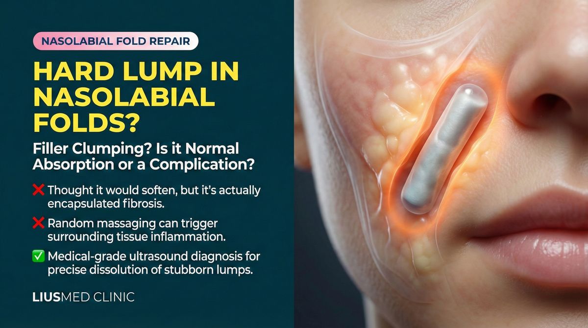
You had filler injected into your nasolabial folds. A few weeks later, you press your finger along the crease and feel something firm beneath the skin—a distinct, cord-like ridge with palpable borders. It wasn't there before. Now you're wondering: is this normal, or has something gone wrong?
Let's break this down systematically.
Classifying Nasolabial Fold Lumps
Not Every "Palpable" Finding Is a Problem
| Type | What It Feels Like | Visible Change | Action Needed |
|---|---|---|---|
| Normal filler texture | Soft, diffuse, compressible with finger pressure | No visible abnormality | None |
| Filler aggregation | More defined cord or beaded shape | May or may not be visible | Case-dependent |
| Fibrous encapsulation | Hard cord, sharp borders, non-compressible | Possible subtle ridge | Recommended |
| Filler displacement | Mass palpable in unexpected location | Poor fold correction; new bulge nearby | Recommended |
| Inflammation/granuloma | Hard, tender, possibly red or swollen | Obvious bump, skin discoloration | Required |
Key Insight: Because the nasolabial fold is a linear structure, filler injected along it is more likely to be palpable than filler placed in most other facial areas. The question is not whether you can feel it, but what it feels like—its consistency, stability, and whether it's causing any problem.
When It's Likely Just Normal Texture
The following situations typically require no intervention:
- You're still within 2–4 weeks post-injection and residual swelling hasn't fully resolved
- The texture feels soft and uniform with no distinct lumps
- It compresses easily under finger pressure
- It doesn't affect facial expression or appearance
- It gradually becomes less noticeable over time
Key Insight: Hyaluronic acid fillers, even when perfectly placed, can sometimes be palpable in areas where skin is thin and the underlying structure is linear. Feeling something doesn't automatically mean something is wrong.
When You Should Seek Evaluation
These signals suggest a visit to a specialist is warranted:
- The lump persists unchanged or worsens beyond 4 weeks post-injection
- The lump is visible—you can see a ridge under certain lighting or angles
- The texture is becoming harder over time rather than softer
- There is tenderness or intermittent pain on palpation
- The lump's location doesn't match where the filler was originally injected (suggesting migration)
- Skin color changes appear over the area
The Diagnostic Value of Ultrasound
High-resolution ultrasound provides critical information in the nasolabial fold region:
- Distribution pattern: Is the filler evenly spread or clumped into deposits?
- Encapsulation status: Has a fibrous capsule formed around the filler?
- Depth assessment: Is the filler at the correct tissue plane, or is it too superficial or too deep?
- Inflammatory reaction: Is there tissue inflammation surrounding the filler?
- Volume estimation: How much residual filler remains and where is it distributed?
This diagnostic clarity is essential because different causes require fundamentally different treatment strategies.
Treatment Strategies by Lump Type
For encapsulated lumps: Dissolving enzymes typically cannot penetrate the fibrous capsule. Ultrasound-guided pinhole extraction provides a more reliable solution.
For filler aggregation without encapsulation: If the filler is hyaluronic acid and not encapsulated, precisely targeted enzyme injection under ultrasound guidance may be effective.
For displaced filler: Ultrasound must first confirm the filler's actual location before an extraction strategy can be designed. Blind treatment risks missing the displaced material entirely.
For inflammatory or granulomatous lumps: Inflammation must be controlled first, then a decision is made about whether the filler needs removal. See lumps appearing years after injection for more detail.
Prevention: Nasolabial Fold Injection Best Practices
- Appropriate product selection: The nasolabial fold requires products with adequate support but not excessive firmness
- Correct depth: Too superficial makes the filler easily palpable; too deep compromises the visible result
- Avoid overfilling: The nasolabial fold has limited tissue space—overfilling increases lump risk significantly
- Even distribution: Injection technique should ensure filler spreads uniformly along the fold rather than accumulating in one spot
If you're concerned about a lump along your nasolabial fold, schedule a consultation. Ultrasound can tell you whether what you're feeling is simply normal filler texture or something that needs attention.
Common questions
Does feeling a cord-like lump along the fold mean the filler went wrong?
Not necessarily. The nasolabial fold is a linear structure, so filler placed here is easier to feel than in most other areas. What matters isn't whether you can feel it, but how it feels — soft and compressible versus hard with sharp borders — and whether it's actually causing a problem. Feeling something on its own isn't a red flag.
How do I know whether to just keep watching it or get it checked?
In the first two to four weeks, if it's soft, even, presses in under your finger and doesn't affect your expression, it's usually residual swelling still settling. Come in for evaluation if it's still firm past four weeks, is getting harder rather than softer, becomes visible under certain lighting, feels tender, or sits somewhere other than where you were injected.
If there's a lump, can't we just dissolve it with enzyme?
It depends on what the lump actually is. If a fibrous capsule has formed around the filler, dissolving enzyme often can't get through that capsule, so it does little. Enzyme has a better chance when it's hyaluronic acid that has clumped without a capsule. That's why we look with ultrasound first and then decide how to handle it.
What can the ultrasound actually show?
It helps us see whether the filler is spread evenly or clumped together, whether a fibrous capsule has formed, what depth it sits at, whether the surrounding tissue is inflamed, and how much is left and where. Since different causes call for different approaches, that picture is what guides the plan.
Is there anything that lowers the chance of a lump in the first place?
A few things help: choosing a product with enough support but not overly firm, placing it at the right depth (too shallow is easy to feel, too deep loses the effect), not overfilling a fold that has limited tissue space, and spreading the filler evenly along the line instead of leaving it in one spot.
Related Reading
- Hard Lump Years After Filler? Don't Rush to Dissolve
- Why Dissolving Enzymes Can't Break Down Your Filler
- Ultrasound-Guided Pinhole Filler Extraction
Related Services

Specialties
Credentials
- Kaohsiung Medical University, School of Medicine
- Attending Physician, Dermatology, Kaohsiung Chang Gung Memorial Hospital
- Attending Physician, Aesthetic Center, Kaohsiung Chang Gung Memorial Hospital
- Visiting Physician, Dermatology, Xiamen Chang Gung Hospital
- Visiting Physician, Aesthetic Center, Xiamen Chang Gung Hospital
"For every surgery, I strive to achieve a good outcome through a small incision and refined technique. Minimally invasive surgery is not just a technique — it's a commitment of respect to every patient."
Recovery after any procedure needs peer support too
Want to learn more?
Schedule a consultation for professional evaluation and advice