Radiesse Complications: Calcium Deposits, Jaw Widening, and Ultrasound-Guided Removal
The Hidden Risk After Radiesse Injection: Why Does the Jaw Keep Getting Wider?
Radiesse has long been a popular filler choice for chin augmentation, nose contouring, and cheek volumization in aesthetic clinics worldwide. Its active ingredient — calcium hydroxylapatite (CaHA) microspheres — is suspended in a carboxymethylcellulose (CMC) gel carrier, delivering immediate volume while stimulating new collagen production. For where Radiesse sits as a material, the filler-revision site has a Radiesse (CaHA) encyclopedia page.
However, an increasing number of patients report troubling changes months or even years after injection: gradual jaw widening, palpable hard lumps, and localized tissue hardening. These are not part of the normal metabolic process — they are the consequence of residual Radiesse triggering tissue fibrosis.
Key Insight: While Radiesse is marketed as being absorbed within 12–18 months, clinical practice reveals that residual deposits and calcification are far from rare, particularly when large volumes are injected or placement depth is incorrect.
Radiesse Composition and Metabolism
Understanding CaHA (Calcium Hydroxyapatite (Aesthefill / Radiesse) — mineral-based filler) Microspheres
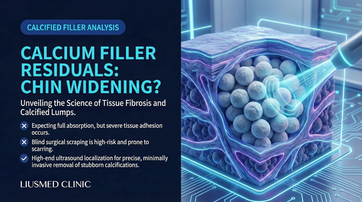
The core component of Radiesse consists of CaHA microspheres approximately 25–45 micrometers in diameter. These particles share the same chemical composition as human bone and teeth minerals. In theory, the body gradually breaks them down into calcium and phosphate ions for natural elimination.
| Component | Function | Expected Metabolism |
|---|---|---|
| CaHA microspheres | Structural support, collagen stimulation | 12–18 months (theoretical) |
| CMC gel carrier | Immediate volume, microsphere distribution | 2–3 months |
| Neocollagen | Sustained volumizing effect | Persists but gradually diminishes |
Why Radiesse Cannot Be Dissolved
Unlike hyaluronic acid fillers, Radiesse has no corresponding dissolving enzyme. Hyaluronidase (enzyme that dissolves HA filler) is designed exclusively to cleave the molecular bonds of hyaluronic acid — it has zero efficacy against CaHA. The occasional claim that hyaluronidase can treat Radiesse complications is pharmacologically unfounded.
Key Insight: No approved medication or enzyme anywhere in the world can dissolve Radiesse. When residual CaHA causes problems, physical extraction remains the only definitive solution.
Common Radiesse Complications
Nodule and Lump Formation
CaHA microsphere aggregation within tissues can produce palpable hard lumps. Contributing factors include:
- Incorrect injection depth: Superficial placement allows microspheres to form visible clusters
- Excessive volume: Over-injection in a single site prevents even microsphere distribution
- Individual tissue response: Some patients mount a more aggressive fibroblastic reaction, accelerating capsule formation
- Poor local circulation: Slower microsphere metabolism leads to prolonged retention
Jaw Widening
The chin is one of the most common Radiesse injection sites, and jaw widening is a particularly distressing complication:
| Cause | Mechanism | Appearance |
|---|---|---|
| Filler spread | Microspheres disperse laterally along fascial planes | Chin appears squared from the side |
| Capsule formation | Fibrous tissue encases the entire deposit | Hard plate palpable on touch |
| Tissue fibrosis | Chronic foreign body reaction stimulates fibrosis | Tight, inelastic chin skin |
| Calcified aggregation | Microspheres cluster into larger calcified masses | Small stone-like feel on palpation |
Tissue Fibrosis (excess scar tissue formation) and Calcification
Long-term CaHA retention can trigger a cascade of pathological changes:
- Foreign body giant cell reaction: Immune cells attempt to engulf microspheres but cannot fully digest them
- Chronic inflammation: Persistent low-grade inflammation stimulates fibroblast activity
- Capsule formation: Fibrous tissue progressively encases the filler deposits
- Dystrophic calcification: Additional calcium deposits may form around retained microspheres
Limitations of Conventional Treatments
Massage and Observation
Many patients are told to "wait — Radiesse will absorb on its own." However, if palpable lumps or contour irregularities persist beyond 18–24 months post-injection, spontaneous absorption is unlikely. By this stage, the capsule has matured, and even if CaHA microspheres eventually degrade, the fibrous capsule will persist.
Steroid Injections
Intralesional steroid injections can temporarily manage inflammatory nodules, but carry significant risks:
| Potential Benefit | Potential Risk |
|---|---|
| Temporary anti-inflammatory effect | Skin atrophy and depression |
| Nodule softening | Hypopigmentation |
| Pain relief | Telangiectasia |
| — | Fat atrophy |
Steroids have limited effect on already-calcified or fibrotic tissue, and repeated injections accumulate tissue damage risk.
Surgical Excision
Traditional open surgical excision can remove Radiesse deposits, but performing open surgery on the face means larger incisions, greater scarring risk, and longer recovery. For patients who initially sought cosmetic enhancement, this is rarely the ideal option.
Ultrasound-Guided Minimally Invasive Extraction: The "See Before You Treat" Strategy
Why Ultrasound Is Critical
Radiesse has a distinctive ultrasound signature — CaHA microspheres appear as hyperechoic (bright white) signals, making ultrasound the ideal tool for locating residual deposits.
| Ultrasound Capability | Clinical Value |
|---|---|
| Hyperechoic identification | CaHA clearly visible on ultrasound, precise localization |
| Depth and extent mapping | Quantifies residual filler distribution |
| Capsule assessment | Evaluates fibrous capsule thickness and extent |
| Vascular mapping | Avoids critical blood vessels |
| Real-time confirmation | Monitors extraction progress throughout the procedure |
The Extraction Procedure
Phase 1: Comprehensive Ultrasound Assessment
- Map all CaHA deposit locations, dimensions, and depths
- Evaluate capsule maturity and degree of fibrosis
- Plan optimal pinhole entry routes
Phase 2: Minimally Invasive Extraction
- Local anesthesia
- Pinhole incision (typically 1–2mm)
- Real-time ultrasound-guided approach to the target area
- Specialized instruments fragment the capsule and extract CaHA deposits
- Continuous ultrasound monitoring confirms extraction progress
Phase 3: Post-Procedure Follow-Up
- Light compression dressing for 24–48 hours
- One-week post-procedure assessment
- Follow-up ultrasound at 1 month and 3 months
Expected Outcomes
| Scenario | Expected Result |
|---|---|
| Single nodule | >90% removal in one session |
| Multiple nodules | May require staged treatment |
| Extensive fibrosis | Significant improvement; multiple sessions possible |
| Jaw widening | Contour gradually restores after removal |
| Injection over 3 years ago | Some CaHA may have degraded; capsule can still be extracted |
Key Insight: The goal of ultrasound-guided extraction is not to remove every microscopic CaHA particle, but to remove the principal deposits causing the clinical problem — restoring both appearance and texture to normal.
Frequently Asked Questions
Will there be a depression after Radiesse removal?
Some degree of volume reduction is normal after extraction. Tissues remodel over 3–6 months. If volume restoration is desired, safe HA (Hyaluronic Acid — sugar molecule naturally in skin, holds water) filler can be placed once healing is complete.
How long after injection can Radiesse be removed?
Extraction is technically possible at any time. Clinically, if significant residual symptoms persist beyond 18 months, active evaluation for extraction is recommended. Earlier treatment means thinner capsules and easier removal.
Will the extraction leave scars?
Pinhole incisions (1–2mm) heal with virtually invisible marks. Compared to traditional surgical excision, the cosmetic advantage of minimally invasive extraction is substantial.
Do Not Let Residual Radiesse Control Your Appearance
If you have experienced jaw widening, hard lumps, or tissue hardening after Radiesse injection — and months or years of waiting have brought no improvement — ultrasound-guided minimally invasive extraction may be the solution you need. The first step is a comprehensive ultrasound evaluation to determine the exact distribution of residual filler.
Further reading:
- Why Dissolving Enzymes Fail When Capsules Form
- Minimally Invasive Filler Lump Extraction Technique
- Lumps Appearing Years After Injection
About the Author
Dr. Ta-Ju Liu
- Current Position: Director, Liusmed Clinic
- Specialties: Minimally invasive surgery, filler complication repair, ultrasound-guided extraction
- Experience: 15+ years of clinical minimally invasive surgery; over 10,000 successful cases
- Philosophy: "Radiesse retention is more common than most realize. Patients should not have to choose between living with lumps and undergoing major surgery — ultrasound-guided minimally invasive extraction offers a third path."

Specialties
Credentials
- Kaohsiung Medical University, School of Medicine
- Attending Physician, Dermatology, Kaohsiung Chang Gung Memorial Hospital
- Attending Physician, Aesthetic Center, Kaohsiung Chang Gung Memorial Hospital
- Visiting Physician, Dermatology, Xiamen Chang Gung Hospital
- Visiting Physician, Aesthetic Center, Xiamen Chang Gung Hospital
"For every surgery, I strive to achieve a good outcome through a small incision and refined technique. Minimally invasive surgery is not just a technique — it's a commitment of respect to every patient."
Recovery after any procedure needs peer support too
Want to learn more?
Schedule a consultation for professional evaluation and advice