Can Facial Lumps Be Surgically Cut Out? Traditional Excision Scarring vs. Minimally Invasive Extraction
"Why Not Just Cut It Out?"
After pharmacological treatments—dissolving enzymes, steroids, 5-FU—have all failed, some practitioners suggest a final option: "Let us just surgically excise the lump." It sounds straightforward and seems like it could solve the problem once and for all. But before deciding, you need to understand the real risks and costs of traditional open surgery on the face.
Traditional surgical excision is a method that works but at a high cost—it can indeed remove the lump, but the accompanying scar, tissue deficit, and recovery period often leave patients trading one problem for another of equal or greater severity.
How Is Traditional Excision Surgery Performed?
Surgical Steps
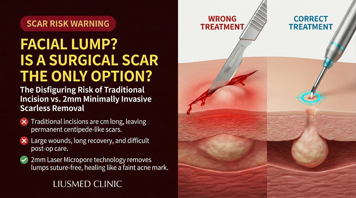
- A skin incision is made over or near the lump (typically 1-3 cm)
- Tissue is separated layer by layer until the lump is reached
- The lump and surrounding tissue are excised together
- The wound is closed with layered sutures
- Post-operative suture removal and wound care
Why Is Open Surgery on the Face Particularly Risky?
| Risk Factor | Description | Severity |
|---|---|---|
| Visible scarring | Any incision on facial skin leaves a permanent scar | High |
| Nerve damage | Incision may sever branches of facial nerves | High |
| Tissue deficit | Excision extent may exceed what is necessary, leaving depressions | Medium-high |
| Asymmetry | Unilateral excision creates side-to-side imbalance | Medium |
| Extended recovery | Facial open surgery requires 2-4+ weeks recovery | Medium |
| Infection risk | Open surgical wounds carry higher infection risk | Medium |
Scarring: The Cruelest Cost of Facial Surgery
Why Are Facial Scars Particularly Conspicuous?
The face is the most scrutinized area in human interaction. Even with the most refined suturing technique, facial incisions will leave some degree of scarring. Scar severity depends on multiple factors:
- Incision placement: Incisions that do not follow relaxed skin tension lines produce more visible scars
- Incision length: The incision needed for lump removal is typically 1-3 cm
- Individual constitution: Patients prone to hypertrophic scarring or keloid formation face worse outcomes
- Post-operative care: Infection or excessive tension worsens scarring
Key Insight: The patient's original concern was "I have an unsightly lump on my face." If the solution leaves a permanent scar, for many patients this is not a true resolution. The goal of treating filler complications should be to restore natural appearance as much as possible—not to replace one problem with another.
The Trap of Over-Excision
In traditional surgery, surgeons habitually ensure "clean margins," often excising a safety border around the lump. In tumor surgery, this is reasonable and necessary. But for filler lumps, this mindset can cause unnecessary tissue sacrifice.
Filler lumps are not tumors—they do not spread or metastasize. Over-excision only creates a larger tissue deficit, and on the face, this deficit directly manifests as depression and asymmetry.
For more analysis on why encapsulated filler cannot rely on dissolution, see: Encapsulation: Why Dissolvers Fail.
Ultrasound-Guided Minimally Invasive Extraction vs. Traditional Open Surgery
| Comparison | Traditional Open Surgery | Ultrasound-Guided Extraction |
|---|---|---|
| Entry size | 1-3 cm incision | Single pinhole (<2mm) |
| Scarring | Permanent visible scar | Nearly invisible |
| Visual guidance | Direct visualization | Real-time ultrasound |
| Surrounding tissue damage | Layer-by-layer dissection required | Precise path preserving normal tissue |
| Recovery time | 2-4 weeks | Several days |
| Anesthesia | May require general or regional | Local anesthesia |
| Material identification | Confirmed only during surgery | Identified before procedure |
| Residual risk | Eyes may miss deep material | Ultrasound confirms clearance |
Key Insight: The reason ultrasound-guided minimally invasive extraction can achieve more thorough filler removal while preserving more normal tissue lies in its ability to see. Ultrasound provides not a blurry outline but precise information about tissue layers, material characteristics, and real-time instrument position.
When Might Traditional Surgery Still Be Necessary?
Objectively, minimally invasive extraction is not appropriate for every situation. In a very small number of scenarios, traditional surgical excision may still be necessary:
- Extremely extensive filler spanning multiple tissue planes: Some historical large-volume injections (such as early unregulated injectable procedures) involve material that has spread extensively across multiple tissue layers
- Severe infection requiring open drainage: Deep abscesses require adequate drainage channels
- Associated tissue necrosis requiring debridement: Areas of established tissue necrosis require thorough debridement
However, even in these situations, ultrasound evaluation remains an indispensable first step—to determine the extent of the problem and formulate a surgical plan.
Core Advantages of Minimally Invasive Technique
The Significance of a Single Pinhole
"One pinhole" is not merely marketing language—it represents a fundamental shift in treatment philosophy:
- Minimized invasion: The entry point is only a pinhole, reducing surface skin damage to the absolute minimum
- Internal precision: Under real-time ultrasound guidance, precise filler extraction is performed through this pinhole
- Tissue integrity preserved: No layer-by-layer dissection and suturing required; surrounding tissue structure is maintained
- Rapid recovery: No swelling and healing burden from a large incision
The Critical Role of Pre-Procedure Ultrasound
Before every extraction procedure, a complete ultrasound scan is performed. This is not merely a "quick look" but the creation of a detailed treatment map:
- Precise filler location, depth, and extent
- Material characteristics and degree of encapsulation
- Pathways of surrounding critical vessels and nerves
- Optimal entry path planning
For more detailed information on the extraction technique, see: Filler Lump Extraction Technique Explained.
Recommendations Before Making a Decision
If you are considering surgical treatment for facial filler lumps, we recommend the following before making a final decision:
- Get an ultrasound evaluation first: Understand the exact material, location, and extent of the lump
- Assess minimally invasive feasibility: The vast majority of cases can be managed with minimally invasive approaches
- Understand scarring risks: If traditional surgery is ultimately needed, fully understand the scar location and severity
- Seek a second opinion: Especially when the first practitioner recommends direct surgical excision
We recommend starting with a comprehensive ultrasound evaluation. Schedule a consultation and let us find the most appropriate treatment plan for your specific situation.
Conclusion
Traditional open surgery is a last resort, not a first choice. In an era when minimally invasive techniques and ultrasound guidance have reached a high level of maturity, the vast majority of facial filler lumps can be safely removed without leaving visible scars. Do not accept a surgery that will leave a permanent mark on your face before you have explored all available options.
Related Services

Specialties
Credentials
- Kaohsiung Medical University, School of Medicine
- Attending Physician, Dermatology, Kaohsiung Chang Gung Memorial Hospital
- Attending Physician, Aesthetic Center, Kaohsiung Chang Gung Memorial Hospital
- Visiting Physician, Dermatology, Xiamen Chang Gung Hospital
- Visiting Physician, Aesthetic Center, Xiamen Chang Gung Hospital
"For every surgery, I strive to achieve a good outcome through a small incision and refined technique. Minimally invasive surgery is not just a technique — it's a commitment of respect to every patient."
Recovery after any procedure needs peer support too
Want to learn more?
Schedule a consultation for professional evaluation and advice