Case Analysis: Vascular Occlusion Emergency Rescue — From Skin Darkening to Recovery
Case Scenario
Patient Background: A woman in her 30s who received hyaluronic acid injections in the nasal and nasolabial fold areas at another clinic. During the injection, the patient began experiencing unusual stinging and burning sensations near the injection site. Approximately 20 minutes after the injection was completed, blanching appeared in the perialar region, which progressed to a violaceous discoloration within one hour.
Emergency Transfer Timeline:
- 20 minutes post-injection: Blanching appeared in the perialar region; patient reported escalating local pain
- 1 hour post-injection: Blanched area began turning purplish-red with slight expansion
- Original clinic attempted pressure and warm compress management, but symptoms continued to worsen
- ~3 hours post-injection: Affected area darkened further with increasingly defined borders
- Original clinic contacted our facility for emergency transfer
- ~5 hours post-injection: Upon arrival at our clinic, the affected area had progressed to deep purple to dark discoloration
Presentation on Arrival:
- Sharply demarcated deep purple to dark skin discoloration from the perialar region to the nasolabial fold
- Noticeably reduced skin temperature in the affected zone
- Patient reported persistent pain and numbness in the affected area
- Capillary refill test indicated severely diminished blood flow in the affected region
- Patient in extreme distress and anxiety
Deep Analysis
Root Cause Analysis
| Aspect | Finding |
|---|---|
| Occlusion mechanism | Filler directly injected into or compressing a branch of the facial artery, interrupting blood flow |
| Affected vessel | Based on ischemic territory distribution, likely involving the alar artery and its branches |
| Time factor | Approximately 5 hours elapsed from symptom onset to rescue initiation — within the critical tissue salvage window |
| Initial ultrasound assessment | Evidence of filler compressing vasculature in the affected zone; significantly diminished blood flow signals |
| Tissue perfusion status | Deep purple to dark discoloration indicating severe ischemia, but not yet progressed to irreversible necrosis |
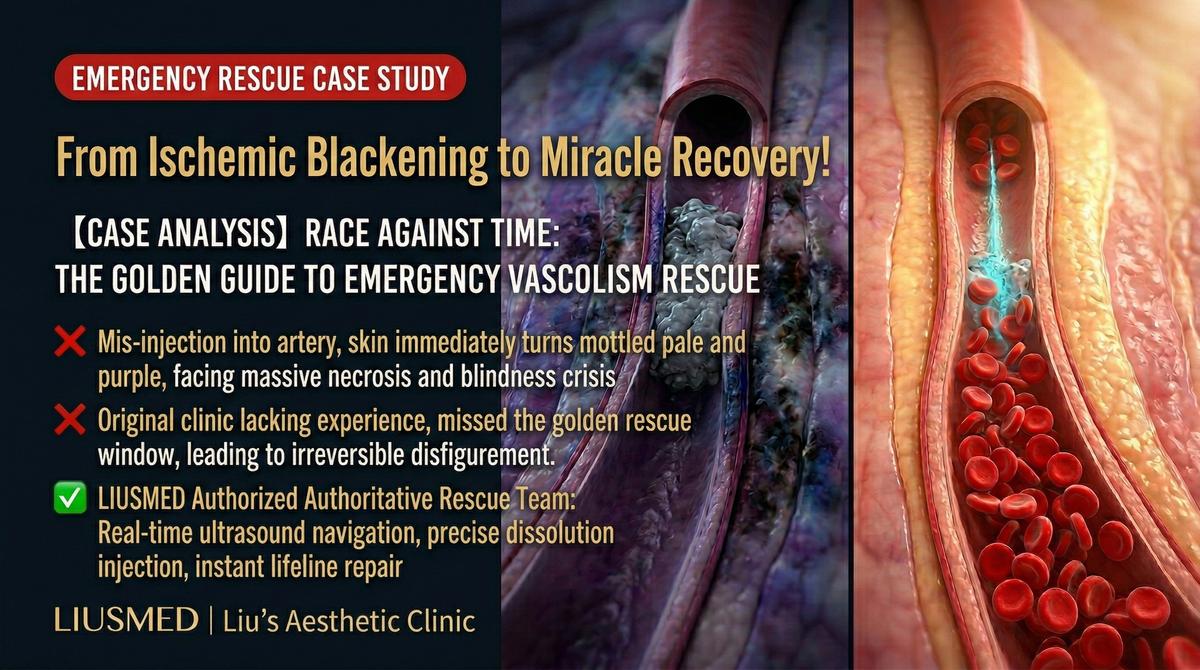
Key Insight: Vascular occlusion is one of the most serious acute complications of filler injection. Time is the critical determinant of tissue survival — the earlier intervention begins, the greater the probability of complete tissue recovery. Classic warning signs include: abnormal pain during injection, blanching or purplish discoloration around the injection area, and decreased skin temperature. Once these signs appear, emergency protocols should be initiated immediately rather than adopting a wait-and-see approach.
Related reading: Vascular Occlusion (filler-induced vascular blockage) Mechanism and Emergency Response
Doctor's Perspective
"The timing of this patient's transfer was critical. While 5 hours is not the ideal intervention window — the earlier the better — it was still within the time frame where tissue salvage remained possible. The deep purple to dark skin color indicated severe ischemia, but through ultrasound assessment, we determined that the tissue had not yet progressed to complete necrosis, meaning a window of opportunity for rescue still existed.
Our emergency strategy was multi-pronged: first, immediately perform ultrasound-guided precision localization to confirm the exact position of the filler and its compression effect on surrounding vasculature; second, administer high-concentration hyaluronidase under ultrasound guidance, delivering it directly to the filler material compressing the vessel; third, implement vasodilation and anticoagulation support measures to facilitate perfusion recovery. The entire rescue process demands both speed and precision — every minute matters for tissue survival."
Treatment Plan and Process
Emergency Strategy
| Phase | Treatment | Timeline |
|---|---|---|
| Phase 1 | Ultrasound emergency assessment and precision localization | Within 15 minutes of arrival |
| Phase 2 | Ultrasound-guided precision hyaluronidase injection | Immediately following assessment |
| Phase 3 | Vasodilation and perfusion support treatment | Concurrent with hyaluronidase |
| Phase 4 | Continuous monitoring and supplemental treatment | 24-48 hours post initial intervention |
Emergency Rescue Process
- Ultrasound emergency assessment: Rapid scanning to confirm filler location, extent of vascular compression, and residual blood flow status
- Precision hyaluronidase injection: Under real-time ultrasound guidance, high-concentration hyaluronidase delivered directly to the filler compressing the vessel
- Sequential supplemental injections: Based on real-time ultrasound monitoring, additional hyaluronidase administered to areas of persistent compression
- Vasodilation support measures: Local and systemic blood flow enhancement therapy implemented concurrently
- Real-time monitoring: Skin color changes and capillary refill assessed every 15-30 minutes
- Follow-up treatment: 24-hour follow-up evaluation to determine need for additional intervention
Rescue Outcome
Approximately 2-3 hours after the initial emergency intervention, the affected skin color began transitioning from dark to a deep red, indicating blood flow restoration. At the 24-hour follow-up, most of the area had progressed to red to light red, with skin temperature gradually returning to normal. Through several days of intensive monitoring and supportive treatment, tissue perfusion progressively normalized.
Key Patient Notes
Special Characteristics of Vascular Occlusion Emergency
| Characteristic | Explanation |
|---|---|
| Time equals tissue | The earlier intervention begins, the greater the chance of complete tissue recovery |
| Recovery is gradual | After blood flow restoration, tissue repair still requires weeks to months |
| Temporary marks may remain | Skin that experienced severe ischemia may undergo a post-inflammatory hyperpigmentation phase; most improve gradually |
| Intensive follow-up is essential | The first few days after rescue require frequent follow-up visits to monitor progress |
| Psychological support matters equally | Patients who experience an occlusion event often suffer significant psychological trauma and need thorough support and communication |
Recovery Timeline
| Timeline | Expected Condition |
|---|---|
| Hours after rescue | Skin color transitions from dark to deep red, indicating blood flow restoration |
| Days 1-3 post-rescue | Continued improvement; affected area may exhibit swelling and inflammatory response |
| Weeks 1-2 post-rescue | Acute phase resolves; tissue enters repair phase; possible hyperpigmentation |
| Months 1-3 post-rescue | Tissue continues to heal; hyperpigmentation gradually improves |
| Month 6 post-rescue | Most cases achieve good recovery; assessment of whether cosmetic follow-up treatment is needed |
Key Insight: Successful vascular occlusion rescue does not mean immediate return to normal. Tissue that has experienced ischemia requires time to repair, and the recovery process may include temporary manifestations such as hyperpigmentation and texture changes. The important thing is to maintain regular follow-up so the physician can assess recovery progress and provide further restorative treatment at the appropriate time.
Clinical Takeaways
- Timely recognition of warning signs is life-saving — abnormal pain during injection and blanching or purplish discoloration are early occlusion signals requiring immediate action
- The time window determines outcomes — the first 6 hours after occlusion onset represent the golden period for rescue
- Ultrasound's value in emergency rescue is irreplaceable — precise localization of the compression source enables precise decompression
- Multi-pronged rescue strategy is essential — hyaluronidase, vasodilation, and perfusion support must proceed simultaneously
- Injection safety is fundamental — understanding the vascular anatomy of facial danger zones is the first step in occlusion prevention
If you experience abnormal symptoms after injection, contact a medical professional immediately. For emergency evaluation, contact our clinic for urgent assistance.
Related reading:
- Vascular Occlusion Mechanism and Emergency Response
- Facial Injection Danger Zones
- Filler Repair Evaluation Process
Related Services

Specialties
Credentials
- Kaohsiung Medical University, School of Medicine
- Attending Physician, Dermatology, Kaohsiung Chang Gung Memorial Hospital
- Attending Physician, Aesthetic Center, Kaohsiung Chang Gung Memorial Hospital
- Visiting Physician, Dermatology, Xiamen Chang Gung Hospital
- Visiting Physician, Aesthetic Center, Xiamen Chang Gung Hospital
"For every surgery, I strive to achieve a good outcome through a small incision and refined technique. Minimally invasive surgery is not just a technique — it's a commitment of respect to every patient."
Recovery after any procedure needs peer support too
Want to learn more?
Schedule a consultation for professional evaluation and advice