Vascular Occlusion Mechanism: The Golden Window from Blanching to Necrosis
The Emergency That Should Never Happen but Must Be Prepared For
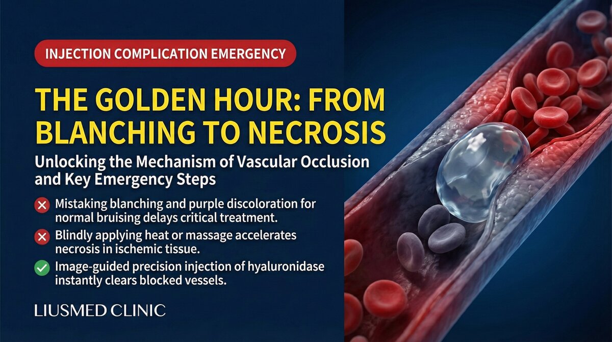
Vascular occlusion is the most feared complication of filler injection. When filler enters an artery or compresses a vessel causing blood flow interruption, downstream tissue can undergo irreversible necrosis within hours. In the most extreme cases, if the occlusion affects the ophthalmic artery, permanent blindness can result.
Understanding this process is not meant to create panic, but to serve a practical purpose: the earlier you recognize signs of occlusion, the better the chance of saving tissue.
Key Insight: Vascular occlusion rescue is a race against time. From skin blanching (ischemia sign) to tissue necrosis (irreversible damage), the window may be only 4-6 hours — even shorter in some areas. Every minute of delay means more tissue that cannot be saved.
Pathophysiology of Vascular Occlusion (filler-induced vascular blockage)
Two Occlusion Mechanisms
Direct intravascular embolism: The needle tip penetrates the arterial wall, and filler enters the arterial lumen under injection pressure, physically blocking blood flow downstream.
Extravascular compression: Large filler volumes compress vessels externally. When external pressure exceeds intravascular pressure, blood flow is blocked.
| Comparison | Direct Arterial Embolism | Extravascular Compression |
|---|---|---|
| Mechanism | Filler enters vessel lumen | Filler compresses vessel externally |
| Onset speed | Immediate or within seconds | Minutes to hours |
| Ischemia distribution | Along arterial territory | Local compression area |
| Severity | Usually more severe | Depends on compression degree |
| Treatment focus | Dissolve intravascular filler | Decompress (remove/disperse filler) |
| Blindness risk | Present (if ophthalmic artery affected) | Very low |
From Blanching to Necrosis (irreversible cell/tissue death): The Clinical Timeline
Phase 1: Blanching (0-30 Minutes)
Blood flow is blocked; downstream tissue suddenly loses oxygen supply. Skin turns pale, with severe disproportionate pain as the key warning sign.
What can be done: Stop injection immediately. For HA (Hyaluronic Acid — sugar molecule naturally in skin, holds water) fillers, inject large volumes of hyaluronidase. Apply warm compresses. Administer oral aspirin. Apply nitroglycerin paste topically.
Phase 2: Ischemic Progression (30 Minutes-6 Hours)
Tissue begins anaerobic metabolism. Lactate accumulates. Cell membranes fail. Blanching areas gradually turn dark purple or blue-gray. Pain intensifies. Blisters may appear.
Phase 3: Tissue Necrosis (6-24 Hours)
Beyond the critical ischemia threshold, cells undergo irreversible death. Skin turns deep purple or black. Eschar forms.
Phase 4: Demarcation and Repair (Days-Weeks)
Necrotic tissue separates from viable tissue. Debridement, wound care, and reconstruction may be needed.
Emergency Rescue Principles
Time Is Tissue
| Time Window | Tissue Status | Salvage Chance |
|---|---|---|
| 0-30 minutes | Ischemic but reversible | High (>80%) |
| 30 min-2 hours | Worsening ischemia | Moderate (50-80%) |
| 2-6 hours | Partial cell death beginning | Limited (20-50%) |
| 6-12 hours | Extensive necrosis progressing | Very low (<20%) |
| >12 hours | Irreversible necrosis | Damage control only |
Key Insight: Blindness is the most irreversible outcome. Once filler reaches the central retinal artery, even immediate treatment has very low probability of restoring vision. Prevention — through ultrasound guidance, avoiding danger zones, slow small-volume injection — is the most important strategy. Learn about facial anatomy danger zones.
Special Area Risks
Nasal Occlusion
The nose is one of the most common sites for filler-related vascular occlusion. Patients with nose filler displacement may have experienced mild vascular compression events without realizing it.
Ophthalmic Artery Occlusion and Blindness
The most catastrophic outcome: retrograde embolization to the ophthalmic artery. The retina is extremely ischemia-sensitive — central retinal artery occlusion can cause permanent vision loss within 60-90 minutes.
The Importance of Prevention
Occlusion rescue is never as effective as prevention:
- Ultrasound-guided injection: Identify vessel locations in real time
- Understanding facial anatomy danger zones
- Small-volume, slow injection: Reduce injection pressure to lower retrograde embolization risk
- Aspiration test: Aspirate before injection to check needle tip is not intravascular
- Cannula use: When appropriate, to reduce vessel puncture risk
- Emergency drugs ready: Hyaluronidase (enzyme that dissolves HA filler), nitroglycerin always available
Learn about the filler repair evaluation process and our vascular occlusion treatment services. Contact us with any concerns.
Key Insight: Vascular occlusion is a war against time, but the best battle is the one that never begins. "Seeing" vessels under ultrasound before injecting is far better than starting rescue after occlusion occurs. Prevention is always more effective than treatment.

Specialties
Credentials
- Kaohsiung Medical University, School of Medicine
- Attending Physician, Dermatology, Kaohsiung Chang Gung Memorial Hospital
- Attending Physician, Aesthetic Center, Kaohsiung Chang Gung Memorial Hospital
- Visiting Physician, Dermatology, Xiamen Chang Gung Hospital
- Visiting Physician, Aesthetic Center, Xiamen Chang Gung Hospital
"For every surgery, I strive to achieve a good outcome through a small incision and refined technique. Minimally invasive surgery is not just a technique — it's a commitment of respect to every patient."
Recovery after any procedure needs peer support too
Want to learn more?
Schedule a consultation for professional evaluation and advice