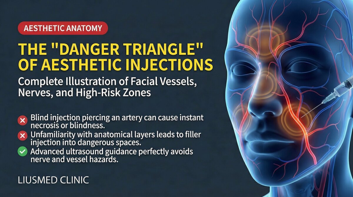
Facial Anatomy: A Complete Guide to Blood Vessels, Nerves, and Injection Danger Zones
Every Needle Passes Through a Complex Anatomical Maze
Filler injection appears simple — disinfect the skin, mark injection points, insert needle, inject, done. The entire process may take less than 15 minutes. But beneath this seemingly calm skin surface lies an extraordinarily complex three-dimensional anatomical structure: arteries coursing through multiple layers, veins forming dense networks, and nerve branches distributed like wiring throughout every tissue plane.
With each injection, the needle tip navigates blindly through this structure — unless you can "see" everything beneath the skin.
Key Insight: Facial vascular anatomy shows significant individual variation. The "standard anatomy" in textbooks represents only statistical averages — your blood vessels may course in entirely different positions. This is why even experienced injectors can encounter unexpected vascular occlusions. "Not being able to see" is the root of the risk.
The Basic Architecture of the Facial Vascular System
Major Arterial Sources
The facial blood supply comes primarily from two arterial systems:
External carotid artery system:
- Facial artery: Ascends along the mandibular border, passing the oral commissure and nasal ala, reaching the medial canthus (becoming the angular artery)
- Superior and inferior labial arteries: Branch from the facial artery, forming circular anastomoses in the lips
- Superficial temporal artery: Supplies the temple and lateral forehead
Internal carotid artery system:
- Ophthalmic artery: Forms extensive anastomotic networks with facial vessels through intraorbital branches
- Supratrochlear and supraorbital arteries: Supply the central and upper forehead
The Dangerous Anastomotic Network
The most critical anatomical fact: extensive anastomoses exist between the external and internal carotid artery systems. This means filler that accidentally enters a facial artery can theoretically travel retrograde through these networks to reach the ophthalmic artery, causing vascular occlusion or even blindness.
| Danger Zone | Vessels Involved | Primary Risk | Risk Level |
|---|---|---|---|
| Glabella | Supratrochlear, supraorbital arteries | Skin necrosis, blindness | Very high |
| Nose | Dorsal nasal, lateral nasal arteries | Skin necrosis, blindness | Very high |
| Nasolabial fold | Facial artery, angular artery | Skin necrosis | High |
| Temple | Superficial temporal artery branches | Skin necrosis, stroke | High |
| Tear trough/infraorbital | Infraorbital artery, angular artery | Skin necrosis, vision impairment | High |
| Forehead | Supratrochlear, supraorbital arteries | Skin necrosis | Medium-high |
| Nasolabial folds | Facial artery | Skin necrosis | Medium |
| Lips | Superior/inferior labial arteries | Tissue necrosis | Medium |
Detailed Analysis of High-Risk Zones
The Nose: One of the Most Dangerous Injection Areas
Nasal filler is the most popular non-surgical rhinoplasty method and simultaneously one of the highest areas for vascular occlusion.
Anatomical risk factors:
- The dorsal nasal artery is a terminal branch of the ophthalmic artery that anastomoses with the angular artery of the facial artery
- Dense lateral nasal artery branches around the nasal ala
- Thin nasal skin with minimal subcutaneous tissue allows fillers to easily compress vessels
- Individual vascular variation is particularly prominent in the nose
Nasal filler displacement and complications are common clinical problems. Learn more about nose filler displacement.
The Glabella: A Highway to the Eyes
The supratrochlear and supraorbital arteries in the glabellar region connect directly to the ophthalmic artery. Filler entering these vessels can travel retrograde extremely rapidly to the central retinal artery, causing irreversible blindness.
The Temple: Hidden Risk Zone
Temple filler improves hollowing, but the superficial temporal artery and deep temporal arteries form complex vascular networks in this area. Deep injection may affect vascular branches leading to the meninges.
The Mechanism of Vascular Occlusion (filler-induced vascular blockage)
From Injection to Onset
When filler accidentally enters an artery or compresses an external vessel:
- Mechanical obstruction: Filler material physically blocks blood flow
- Vasospasm: Vessel walls contract violently when stimulated, further reducing blood flow
- Retrograde embolization: Under injection pressure, filler may travel against blood flow direction to reach distant branches
- Secondary thrombosis: After blood flow stasis, clotting factors activate, forming thrombi that expand the blockage area
Key Insight: Vascular occlusion is a medical emergency. From the appearance of skin blanching and pain to tissue necrosis, there may be only hours in the rescue window. Learn more about the golden time for vascular occlusion emergency treatment.
Nerve Distribution and Injection Risks
Beyond vascular risks, facial nerve distribution must also be well understood:
Three Branches of the Trigeminal Nerve
- Ophthalmic nerve (V1): Supplies sensation to forehead and upper eyelid
- Maxillary nerve (V2): Supplies sensation to mid-face (cheek, nasal ala, upper lip)
- Mandibular nerve (V3): Supplies sensation to lower face and motor function to masticatory muscles
Facial Nerve Branches
The facial nerve controls facial expression muscles. Its branches are particularly superficial in the preauricular and buccal regions, making them vulnerable to injection-related injury that may cause temporary or permanent facial asymmetry.
| Nerve Injury Type | Symptoms | Recovery Time |
|---|---|---|
| Temporary sensory numbness | Decreased sensation at injection site | Usually weeks to months |
| Temporary motor injury | Local expression muscle weakness | Usually weeks |
| Permanent sensory injury | Persistent abnormal sensation | May not fully recover |
| Permanent motor injury | Permanent facial asymmetry | May require surgical repair |
How Ultrasound Changes Injection Safety
"Seeing" Beneath the Skin
High-resolution ultrasound provides real-time anatomical information before, during, and after injection:
Pre-injection assessment: Personalized vascular mapping, identification of variant arteries, confirmation of prior filler residue locations, and tissue layer evaluation.
Intra-procedural guidance: Real-time needle tip confirmation, avoidance of identified vessels, monitoring of filler spread direction, and early detection of abnormalities.
Post-injection monitoring: Confirmation of final filler distribution, assessment for signs of vascular compression, and baseline data for future adjustments.
| Comparison | Blind Injection | Ultrasound-Guided |
|---|---|---|
| Vessel identification | Relies on anatomical knowledge | Real-time visualization |
| Individual variation | Cannot confirm | Can identify |
| Filler localization | Tactile judgment | Precise positioning |
| Old residue detection | Impossible | Detectable |
| Early complication detection | Difficult | May detect before symptoms |
Common questions
Why can filler injection sometimes cause blindness?
Because the two arterial systems in the face — the external and internal carotid — are linked by many connecting channels called anastomoses. If filler accidentally gets into an artery, it can in theory travel backward through those channels toward the ophthalmic artery and cut off blood flow to the eye, which in severe cases affects vision or leads to blindness. The glabella and the nose sit especially close to those eye vessels, which is why the risk there is so high.
Which parts of the face carry the highest injection risk?
The article puts the glabella and nose in the very-high-risk group because their vessels connect straight to the eye. The area beside the nostril, the temple, and the tear trough/under-eye are high risk, while the nasolabial folds and lips sit lower. That said, everyone's vessels run a little differently, so these levels are only a rough guide — your own anatomy is what really matters.
After a filler injection, what symptoms mean I should get help immediately?
If the treated skin suddenly turns pale, you feel pain that is out of proportion to normal swelling, or your vision changes, seek medical care right away. Vascular occlusion is an emergency — from skin blanching to tissue death there may be only a few hours to act, so it isn't something to wait out.
What is the difference between ultrasound guidance and blind injection?
With blind injection the doctor judges where the needle tip and vessels are from anatomical knowledge and feel. Ultrasound lets the doctor actually see the vessels and tissue layers under the skin before, during, and after the injection. It can map your individual vessels ahead of time, steer around them, and even pick up old filler left from before — things blind injection simply can't do.
Anatomy textbooks look so clear — why do problems still happen?
Because textbooks show a statistical average, the "typical" face rather than yours. Facial vessels vary a lot from person to person, and yours may run in completely different positions. That's why even an experienced injector working from memorized anatomy can run into trouble — real safety comes from seeing where this particular person's vessels actually are.
Understand Your Risk, Make Informed Choices
Facial anatomical complexity reminds us that filler injection is not a "simple lunchtime procedure." Every needle operates in a three-dimensional space filled with vessels and nerves.
Risk reduction strategies:
- Choose an injector with ultrasound capability for safer decisions
- Understand the risk level of your injection area
- Confirm emergency protocols — does your injector have vascular occlusion emergency plans?
- Know when to seek help — immediate medical attention for blanching, severe pain, or vision changes
Filler migration and complications are closely related to anatomical structures. Learn about how fillers migrate and the complete filler repair evaluation process.
If you are considering filler injection or facing filler-related concerns, contact us for ultrasound assessment. Learn about our vascular occlusion treatment and filler repair services.
Key Insight: The best injection safety strategy is not memorizing "average positions" from anatomy textbooks, but using ultrasound to see in real time where "this person's" vessels actually are. Personalized safety begins with personalized visualization.

Specialties
Credentials
- Kaohsiung Medical University, School of Medicine
- Attending Physician, Dermatology, Kaohsiung Chang Gung Memorial Hospital
- Attending Physician, Aesthetic Center, Kaohsiung Chang Gung Memorial Hospital
- Visiting Physician, Dermatology, Xiamen Chang Gung Hospital
- Visiting Physician, Aesthetic Center, Xiamen Chang Gung Hospital
"For every surgery, I strive to achieve a good outcome through a small incision and refined technique. Minimally invasive surgery is not just a technique — it's a commitment of respect to every patient."
Recovery after any procedure needs peer support too
Want to learn more?
Schedule a consultation for professional evaluation and advice