Special Considerations for Forehead and Temple Filler Extraction
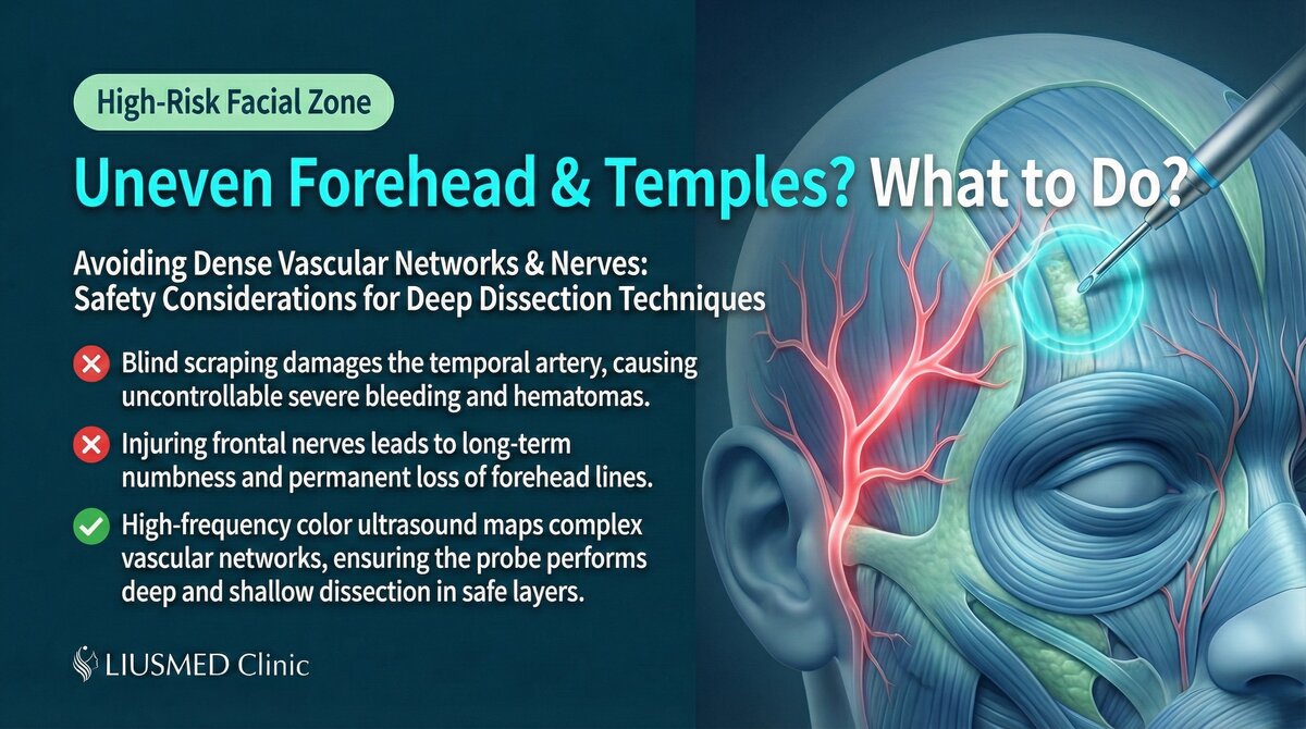
Forehead and Temples: High-Risk Revision Zones
The forehead and temples have seen rapid growth in filler volume in recent years, but they are also among the highest-risk areas for revision. These zones share common characteristics: rich deep vasculature, low surface visibility, and elevated filler migration risk.
Anatomical Specifics
| Structure | Forehead | Temple |
|---|---|---|
| Skin thickness | Moderate | Thinner |
| Major arteries | Supraorbital, supratrochlear | Superficial temporal artery (STA) and branches |
| Major nerves | Supraorbital, supratrochlear | Temporal branch of facial nerve |
| Common filler planes | Subcutaneous to supraperiosteal | Superficial temporal fascia to deep temporal fascia |
| Migration direction | Downward to glabella, lateral to temples | Downward to zygomatic arch |
| Surgical risk level | Medium-High | High |
Key Insight: The temple is one of the highest-risk zones for filler revision on the entire face. The superficial temporal artery runs superficially with dense branching, and any surgical maneuver must confirm vessel position under ultrasound guidance.
Common Forehead Filler Problems
| Problem | Presentation | Common Cause |
|---|---|---|
| Migration to glabella | Glabellar elevation or irregularity | Gravity and muscle contraction |
| Surface irregularity | Uneven forehead contour | Inconsistent injection depth |
| Lump formation | Palpable firm nodule | Filler aggregation or fibrosis |
| Lateral migration | Spread from forehead center toward temples | Natural flow of liquid filler |
For more on forehead filler migration, see Forehead Filler Migration and Bumps.
Special Risks of Temple Filler
The Critical Role of the Superficial Temporal Artery
| Feature | Description |
|---|---|
| Location | On the surface of or within the superficial temporal fascia |
| Course | Curves upward from the preauricular area toward the vertex |
| Branches | Frontal and parietal branches |
| Ultrasound appearance | Pulsatile tubular structure with bright color Doppler signal |
| Surgical significance | Injury can cause major hemorrhage difficult to control with compression |
Temple's Unique Fascial Architecture
The temple has a unique layered structure unlike other facial areas:
- Skin
- Subcutaneous tissue (containing STA, temporal branch of facial nerve)
- Superficial temporal fascia
- Superficial temporal fat pad
- Deep temporal fascia
- Temporalis muscle
Filler may reside in any of these layers, and different materials tend to distribute at different planes.
Ultrasound-Guided Extraction Strategies
Forehead Extraction Key Points
| Step | Operation | Precaution |
|---|---|---|
| Scanning | Complete scan from forehead to glabella | Confirm any migration |
| Vessel marking | Mark supraorbital artery position | Use color Doppler |
| Incision design | Hidden at hairline or glabella | Select most concealed location |
| Extraction | Layer-by-layer, superficial to deep | Continuous ultrasound monitoring |
| Verification | Ultrasound confirmation of clearance | Watch for deep supraperiosteal residual |
Temple Extraction Key Points
| Step | Operation | Precaution |
|---|---|---|
| Scanning | Complete scan from temple to zygomatic arch | Track STA course |
| STA marking | Precisely mark main trunk and branches | Keep color Doppler continuously active |
| Incision design | Hidden within the hairline | Maintain safe distance from STA |
| Extraction | Extremely cautious layered operation | Full-procedure color Doppler monitoring |
| Hemostasis check | Careful post-op confirmation of no bleeding | Temple hematoma has serious consequences |
Key Insight: Temple extraction safety depends entirely on ultrasound real-time guidance. In this zone, operating without ultrasound is effectively blind surgery — an unacceptable risk.
Post-Dissolution Temple Hollowing
Some patients develop severe hollowing after attempting to dissolve temple HA (Hyaluronic Acid — sugar molecule naturally in skin, holds water). See Temple Hollowing After Dissolving for more.
Conclusion: High-Risk Areas Require the Highest Safety Standards
Forehead and temple filler extraction demands thorough understanding of deep anatomy combined with ultrasound guidance for safety. If you have filler concerns in these areas, choose a physician with ultrasound capability and extensive experience.
Contact Liusmed Clinic to arrange a consultation. Related reading: Filler Lump Extraction Technique
A staged real example: two years of Ellansé forehead lumps, from ultrasound map to three months after repair.
Related Services

Specialties
Credentials
- Kaohsiung Medical University, School of Medicine
- Attending Physician, Dermatology, Kaohsiung Chang Gung Memorial Hospital
- Attending Physician, Aesthetic Center, Kaohsiung Chang Gung Memorial Hospital
- Visiting Physician, Dermatology, Xiamen Chang Gung Hospital
- Visiting Physician, Aesthetic Center, Xiamen Chang Gung Hospital
"For every surgery, I strive to achieve a good outcome through a small incision and refined technique. Minimally invasive surgery is not just a technique — it's a commitment of respect to every patient."
Recovery after any procedure needs peer support too


Want to learn more?
Schedule a consultation for professional evaluation and advice