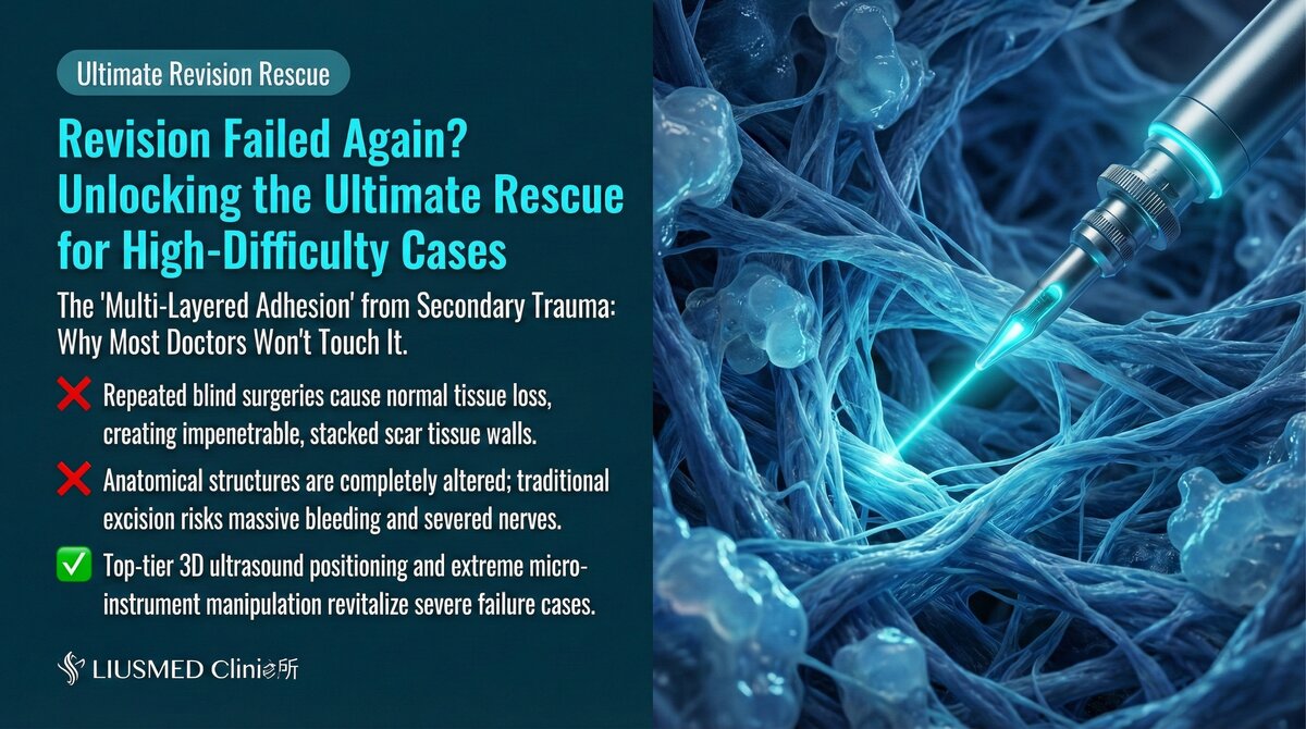
The Complexity of Secondary Revision: Why Failed Repairs Are Harder to Fix
Why Is "Revising a Revision" So Difficult?
When a filler revision surgery fails to achieve the expected outcome — or even creates new problems — the patient needs a secondary revision. This is widely recognized as the most challenging work in the filler revision field.
The reason is straightforward: the first revision has already altered the original tissue architecture. Fibrosis (excess scar tissue formation) is more severe, anatomical landmarks may have been destroyed, and scar tissue has obscured once-clear tissue planes — all of this causes secondary revision difficulty to increase exponentially.
Key Insight: Secondary revision difficulty is not twice that of primary revision — it may be several times greater. Every surgery leaves traces in the tissue, and these traces progressively narrow the operating space and raise the risk profile for each subsequent procedure.
Unique Challenges of Secondary Revision
Tissue-Level Changes
| Change | Impact | Increased Risk |
|---|---|---|
| Worsened fibrosis | Tissue becomes hard, loses elasticity | Extraction difficulty increases |
| Scar formation | Surgical pathways blocked by scar | New operative routes must be found |
| Vascular changes | Original vessels may be damaged or displaced | Bleeding risk increases |
| Neural changes | Nerve pathways may be altered | Nerve injury risk increases |
| Plane obliteration | Previously clear anatomical planes destroyed | Precise operation becomes more difficult |
| Tissue deficiency | Depressions from over-extraction | Reconstruction becomes more complex |
Filler-Level Changes
| Change | Description |
|---|---|
| Residual filler | Portions not fully removed in the first surgery |
| Fragmentation | Filler becomes scattered after partial removal |
| Deep displacement | Surgical manipulation may push filler to deeper layers |
| Mixed materials | Different materials from different injection sessions may coexist |
| Thicker encapsulation | Residual filler enclosed by denser fibrous tissue |
Common Types of Revision Failure
Why Does the First Revision Fail?
| Failure Type | Cause | Outcome |
|---|---|---|
| Incomplete clearance | No ultrasound guidance; deep residuals missed | Problem persists or recurs |
| Over-extraction | Excessive removal causing tissue deficiency | Depression, asymmetry |
| New iatrogenic damage | Vascular or nerve injury during operation | New complications |
| Wrong plane | Operating in the incorrect tissue layer | Normal tissue destroyed |
| Improper dissolution | Non-selective dissolution affecting normal tissue | Depression, unevenness |
| Infection | Surgical site infection | Further tissue damage |
Key Insight: Most revision failures trace back to two root causes: operating without ultrasound guidance or insufficient revision experience. Operating without visualization is effectively blind surgery.
Ultrasound Assessment for Secondary Revision
Why Secondary Revision Needs Ultrasound Even More
In tissue already altered by surgery, the importance of ultrasound is amplified to its maximum:
| Assessment Need | Information Ultrasound Provides |
|---|---|
| Residual filler | Precise location of filler missed in the first surgery |
| Fibrosis extent | Evaluation of fibrosis range and severity |
| Scar distribution | Confirmation of scar tissue location and extent |
| Current vascular status | Verification of post-surgical vessel courses |
| Tissue deficiency | Assessment of depression from over-extraction |
| Normal tissue | Identification of remaining normal tissue structures |
Pre-Operative Ultrasound Assessment Workflow
| Step | Content | Purpose |
|---|---|---|
| Complete scan | Full scan of surgical area and surroundings | Establish comprehensive current status map |
| Residual localization | Mark positions of residual filler | Plan extraction targets |
| Fibrosis assessment | Evaluate depth and extent of fibrosis | Estimate extraction difficulty |
| Vascular remapping | Re-confirm vessel courses | Update safety roadmap |
| Comparative assessment | Compare with contralateral side or pre-op images | Set revision goals |
| Feasibility judgment | Comprehensive assessment of surgical viability | Determine suitability for re-operation |
Surgical Strategy for Secondary Revision
Strategic Differences from Primary Revision
| Strategy Item | Primary Revision | Secondary Revision |
|---|---|---|
| Incision choice | Optimal location available | May be limited by prior scars |
| Operating space | Relatively ample | Compressed by fibrosis |
| Extraction difficulty | Standard | Significantly increased |
| Bleeding risk | Standard | Increased (altered vessel courses) |
| Conservative approach | Standard conservative | Even more conservative |
| Staged strategy | As needed | Strongly recommended |
| Ultrasound dependency | High | Extremely high |
Key Surgical Execution Points
- Maximum conservative principle: Better to leave a small residual than risk damaging normal tissue
- Multi-session staged strategy: Nearly all secondary revisions should be divided into 2–3 sessions
- Full-procedure ultrasound guidance: Every operative step performed under ultrasound monitoring
- Real-time strategy adjustment: Immediate strategy modification based on intraoperative findings
- Sufficient recovery intervals: Allow adequate time between sessions for tissue recovery
Key Insight: The golden rule of secondary revision is "small amounts, multiple sessions." Aggressive operation in already-damaged tissue only creates more damage. Staged extraction gives tissue time to recover and allows the physician to reassess between each session.
Managing Severe Fibrosis
One of the most common challenges in secondary revision is severe fibrosis. For more on fibrosis management, see Severe Adhesion and Fibrosis Extraction.
| Fibrosis Severity | Management Strategy | Expected Outcome |
|---|---|---|
| Mild | Meticulous separation then extraction | Good |
| Moderate | Requires longer time and more sessions | Acceptable |
| Severe | Partial extraction + long-term follow-up | May need to accept some residual |
| Extreme | Conservative observation primarily | Symptom improvement as goal |
Patient Expectation Management
Realistic Expectations for Secondary Revision
| Aspect | Realistic Expectation | Unrealistic Expectation |
|---|---|---|
| Complete clearance | Significant improvement with possible small residual | 100% removal of all filler |
| Appearance recovery | Noticeable improvement with possible minor imperfections | Return to perfect pre-injection state |
| Recovery time | Longer than primary revision | Same as primary revision |
| Number of sessions | May require 2–3 sessions | Resolving everything in one session |
| Final results | Assessed at 3–6 months | Immediately visible final outcome |
How to Avoid Needing Secondary Revision
Keys to Successful Primary Revision
- Choose a physician with ultrasound capability: Ultrasound guidance dramatically reduces revision failure rates
- Complete pre-operative assessment: Thorough understanding of the problem enables correct surgical planning
- Experienced revision specialist: The learning curve for revision surgery is steep
- Realistic expectation setting: Thorough communication with your physician about anticipated outcomes
- Proper post-operative care: Following physician instructions for aftercare
Post-Operative Care and Follow-Up
| Timeline | Special Considerations |
|---|---|
| Weeks 1–2 | Stricter care period, longer than primary revision |
| Month 1 | Evaluate initial recovery; decide if next session needed |
| Month 3 | Interim assessment; tissue beginning to stabilize |
| Month 6 | Evaluate final results; develop follow-up plan |
| Year 1 | Long-term follow-up to confirm stability |
Conclusion: Secondary Revision Demands the Highest Level of Expertise
Secondary revision is the most challenging surgery in the filler revision field. Altered tissue architecture, more severe fibrosis, lost anatomical landmarks — all of this demands the highest caliber of ultrasound interpretation ability and surgical skill from the physician.
If you have experienced a failed revision or are dissatisfied with your current revision outcome, Liusmed Clinic has the full capability to handle these highly complex cases.
Contact Liusmed Clinic to arrange a detailed evaluation.
Related reading: Severe Adhesion and Fibrosis Extraction, Filler Lump Extraction Technique, Filler Repair Evaluation Process
Related Services

Specialties
Credentials
- Kaohsiung Medical University, School of Medicine
- Attending Physician, Dermatology, Kaohsiung Chang Gung Memorial Hospital
- Attending Physician, Aesthetic Center, Kaohsiung Chang Gung Memorial Hospital
- Visiting Physician, Dermatology, Xiamen Chang Gung Hospital
- Visiting Physician, Aesthetic Center, Xiamen Chang Gung Hospital
"For every surgery, I strive to achieve a good outcome through a small incision and refined technique. Minimally invasive surgery is not just a technique — it's a commitment of respect to every patient."
Recovery after any procedure needs peer support too
Want to learn more?
Schedule a consultation for professional evaluation and advice