Severe Tissue Adhesion from Multiple Procedures — Can Minimally Invasive Surgery Still Achieve Clean Removal?
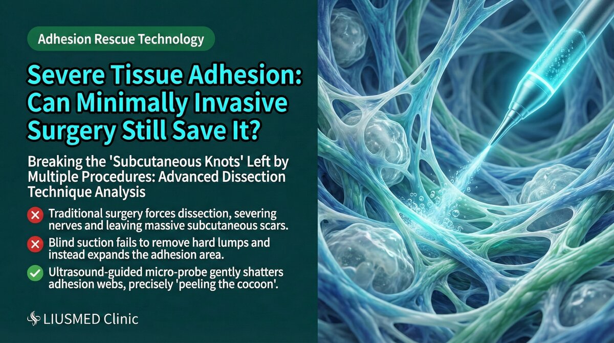
Why Do Multiple Procedures Cause Severe Adhesion?
Every filler injection or revision surgery creates some degree of micro-trauma to local tissue. The body's wound healing mechanism generates fibrous tissue (scar tissue) after each injury. When this process repeats, fibrous tissue accumulates progressively, ultimately forming severe tissue adhesion and fibrosis.
The Adhesion Formation Process
| Stage | Tissue Change | Clinical Presentation |
|---|---|---|
| First injection | Mild foreign body reaction | Usually no apparent problem |
| Multiple injections | Repeated stimulation, fibrous tissue accumulates | Firm texture, surface irregularity |
| Dissolving/repair attempts | Additional trauma accelerates fibrosis | Tissue layer disruption |
| Multiple failed repairs | Extensive fibrosis and adhesion | Tissue stiffness, deformity, pain |
| Severe adhesion stage | Normal tissue layers completely obliterated | Skin, subcutaneous tissue, fascia fused into one mass |
Key Insight: Each unsuccessful repair attempt adds new fibrosis to already damaged tissue. This is like repeatedly creating new wounds on an existing wound — scarring only worsens. Therefore, the quality of repair matters far more than the number of attempts.
Adhesion on Ultrasound
Normal Tissue vs. Adhesed Tissue
| Ultrasound Feature | Normal Tissue | Adhesed Tissue |
|---|---|---|
| Layer structure | Skin, subcutaneous fat, fascia clearly stratified | Layers blurred or completely obliterated |
| Echo characteristics | Each layer has distinct echo properties | Uniform hyperechoic appearance suggesting fibrosis |
| Mobility | Layers glide freely between each other | Layers fused and fixed, no sliding |
| Vascular distribution | Normal vessel course | Vessels may be encased or displaced by fibrous tissue |
| Filler boundaries | Clear boundaries between filler and tissue | Filler interwoven with fibrous tissue, boundaries unclear |
Adhesion Severity Grading
| Grade | Ultrasound Findings | Surgical Difficulty | Management Strategy |
|---|---|---|---|
| Mild | Localized fibrous bands, layers still identifiable | Moderate | Single-session minimally invasive extraction |
| Moderate | Multiple fibrous bands, some layers obliterated | Higher | Minimally invasive extraction with meticulous dissection |
| Severe | Extensive fibrosis, layers completely obliterated | High | Staged minimally invasive treatment |
| Very severe | Tissue completely encased in fibrosis | Very high | Staged treatment with conservative strategy |
Minimally Invasive Strategies for Severe Adhesion
Unique Advantages of Ultrasound Guidance
In severe adhesion, ultrasound guidance offers advantages over traditional surgery:
- Real-time boundary identification: Even when layers are blurred, ultrasound can still differentiate filler from fibrous tissue
- Safe navigation: Tracking displaced vessels and nerves within adhesed tissue
- Precise dissection: Accurate separation of fibrous bands under direct visualization
- Immediate confirmation: Verification of clearance after each dissection step
Layered Progressive Strategy
Severe adhesion cannot be resolved in a single rush. A "layered progressive" approach is essential:
| Step | Operation | Purpose |
|---|---|---|
| Layer 1 | Begin from outermost (superficial) layer, separate skin from subcutaneous adhesion | Restore skin mobility |
| Layer 2 | Advance into subcutaneous fat layer, separate filler from surrounding fibrosis | Expose the filler body |
| Layer 3 | Extract separable filler | Reduce foreign body burden |
| Layer 4 | Address deep residual | Clear deep-layer filler |
| Assessment | Ultrasound confirmation of clearance | Determine if staged treatment is needed |
Which Fillers Most Commonly Cause Severe Adhesion?
| Filler Type | Adhesion Severity | Reason |
|---|---|---|
| Liquid silicone | Very high | Free migration, continuous foreign body reaction |
| PMMA | Very high | Permanent foreign body, chronic inflammation |
| Sculptra | High | Stimulates extensive collagen proliferation |
| Ellanse | Medium-high | Polycaprolactone component persists long-term |
| Radiesse | Moderate | Granule deposition can stimulate fibrosis |
| Hyaluronic acid | Lower | Biodegradable, but repeated injections can still cause adhesion |
For more on why dissolving agents fail with encapsulated fillers, see Why Dissolvers Fail on Encapsulated Fillers.
The Necessity and Planning of Staged Surgery
Why Can't Everything Be Done in One Session?
Severely adhesed cases often require staged surgery because:
- Operating time limits: Prolonged surgery increases tissue edema and bleeding risk
- Tissue tolerance: Excessive dissection causes additional injury
- Monitoring recovery response: Tissue reaction to surgery needs observation
- Progressive fibrosis reduction: Tissue needs time to remodel between sessions
Typical Staged Surgery Plan
| Session | Interval | Goal |
|---|---|---|
| First | — | Remove primary filler, initial adhesion release |
| Second | 2–3 months | Address residual filler, further dissection |
| Third (if needed) | 2–3 months | Refinement, address deep residual |
| Final assessment | 3–6 months post-op | Confirm final result, evaluate reconstruction need |
Key Insight: Staged surgery is not a technical compromise but a physiologically rational plan. Giving tissue adequate recovery time improves both the efficiency and safety of each subsequent session.
Frequently Asked Questions
"My situation is already very severe — can minimally invasive surgery really help?"
Minimally invasive surgery remains effective in severe adhesion, but results may require cumulative sessions. Setting realistic expectations is important — revision of severe adhesion is a process, not a single event.
"Why did my previous doctor say it couldn't be treated?"
Some physicians may lack experience with severe adhesion cases or may not have ultrasound guidance equipment and capability. Under ultrasound guidance, even in tissue where layers are completely disrupted, differences between filler and fibrous tissue can still be identified, providing surgical navigation.
Conclusion: Adhesion Does Not Mean Untreatable
Severe tissue adhesion certainly increases revision difficulty, but it does not mean the situation is beyond help. Through ultrasound guidance's real-time imaging capability, layered progressive strategy, and rational staged surgery planning, even the most complex adhesion cases can achieve gradual improvement.
If you are facing tissue adhesion problems from multiple cosmetic procedures or failed revisions, contact Liusmed Clinic for an evaluation. For more on extraction techniques, see Filler Lump Extraction Technique and Ultrasound-Guided Pinhole Extraction Explained.
Related Services
Specialties
Credentials
- Kaohsiung Medical University, School of Medicine
- Attending Physician, Dermatology, Kaohsiung Chang Gung Memorial Hospital
- Attending Physician, Aesthetic Center, Kaohsiung Chang Gung Memorial Hospital
- Visiting Physician, Dermatology, Xiamen Chang Gung Hospital
- Visiting Physician, Aesthetic Center, Xiamen Chang Gung Hospital
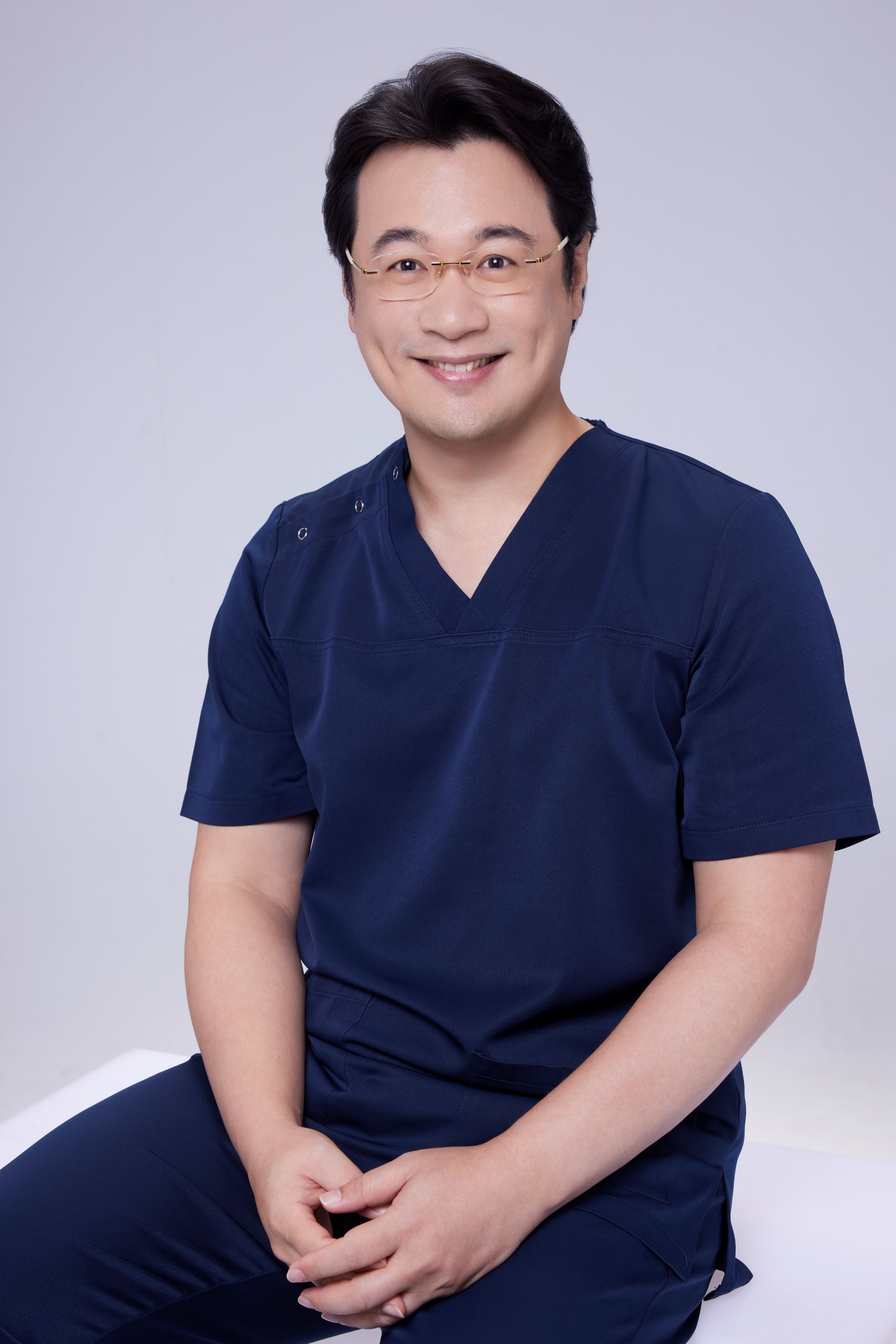
"For every surgery, I strive to achieve a good outcome through a small incision and refined technique. Minimally invasive surgery is not just a technique — it's a commitment of respect to every patient."
Recovery after any procedure needs peer support too

Want to learn more?
Schedule a consultation for professional evaluation and advice