Can High-Resolution Ultrasound Identify Which Filler Is Causing Inflammation?
"What Is Actually Inside?" — The First Question in Repair
There is a lump on your face. Perhaps it was injected three years ago. Perhaps you cannot recall the brand, or are unsure of the material. Perhaps you received different products at different clinics over time. Now there is a problem — swelling, lumps, asymmetry — and you need to know what is inside before deciding how to handle it.
This is where high-resolution ultrasound provides its greatest value. It does not merely tell you "something is there" — it provides critical clues about what that something might be.
Key Insight: Different filler types present different echo characteristics on ultrasound — just as different materials appear different on X-ray. Understanding these characteristic differences is the first step toward precise treatment planning. Blindly treating a lump of unknown composition is like prescribing medication without knowing the diagnosis.
How Ultrasound "Sees" Fillers
Basic Principles
An ultrasound probe emits high-frequency sound waves (typically 15-50 MHz). As waves travel through tissue, they reflect off interfaces between materials of different density. Different materials — with different densities, water content, and particle structures — produce different echo patterns.
Echo Classification
| Echo Type | Ultrasound Appearance | Common Correlates |
|---|---|---|
| Anechoic | Completely black | Fluid (water, pus, liquefactive necrosis) |
| Hypoechoic | Dark gray | Most soft tissue, HA fillers |
| Isoechoic | Medium gray | Material similar to surrounding tissue |
| Hyperechoic | Bright white | Calcification, silicone, certain particulate fillers |
| Strongly hyperechoic with shadowing | Very bright white with posterior dark | Calcified tissue, metal, bone |
Ultrasound Characteristics of Each Filler Type
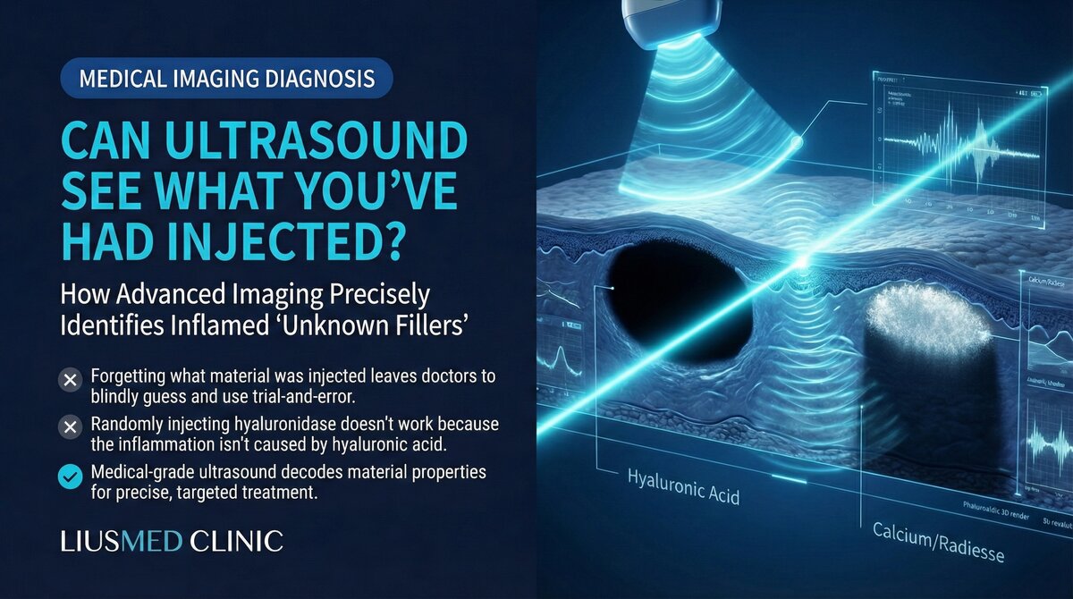
Hyaluronic Acid (HA)
Typical appearance: Hypoechoic to anechoic round or oval deposits with usually clear boundaries. Non-encapsulated HA (Hyaluronic Acid — sugar molecule naturally in skin, holds water) appears as a dark "bubble" on ultrasound.
Changes over time: Fresh injection shows uniform hypoechogenicity with clear borders. After months, heterogeneous echogenicity may appear. After years, residual fragments present as irregular hypoechoic areas possibly surrounded by hyperechoic capsule.
Key distinction: HA is the only filler that responds to hyaluronidase. If ultrasound confirms HA without encapsulation, enzymatic dissolution may be the first-line option. But if already encapsulated, dissolution efficacy is limited.
Poly-L-Lactic Acid (PLLA/Sculptra)
Typical appearance: Hyperechoic bright spots scattered through tissue, resembling a "starry sky" or "snowflake" pattern.
Polycaprolactone (PCL/Ellanse)
Typical appearance: Hyperechoic microspheres within a hypoechoic gel carrier, presenting a "mixed echo" pattern.
Calcium Hydroxylapatite (CaHA/Radiesse)
Typical appearance: Strongly hyperechoic with posterior acoustic shadowing. The calcium component produces very strong echo reflection, making it highly identifiable on ultrasound.
Silicone/Permanent Fillers
Typical appearance: Hyperechoic irregular deposits, often with a "snowstorm" effect — multiple intense echoes obscuring posterior structures.
Autologous Fat
Typical appearance: Isoechoic to hypoechoic, similar to surrounding adipose tissue. Surviving fat is difficult to distinguish from normal fat, but necrotic fat forms oil cysts (anechoic round structures) or calcification.
Key Insight: Each filler type has its own ultrasound "fingerprint." While identification cannot always be made with absolute certainty, an experienced ultrasound operator can make highly accurate assessments in most cases — far superior to blind guessing.
Ultrasound in Diagnosing Filler Complications
Inflammation vs. Infection vs. Granuloma
When swelling occurs at an injection site, ultrasound helps differentiate causes:
| Cause | Ultrasound Features | Doppler Blood Flow |
|---|---|---|
| Simple inflammation | Tissue edema around filler | Mildly increased |
| Acute infection/abscess | Liquefactive necrosis zone (anechoic) | Markedly increased peripherally |
| Biofilm | Heterogeneous echo around filler | Intermittently increased |
| Granuloma | Structured mixed-echo nodule | Increased intranodular flow |
| Capsular contracture | Hyperechoic ring surrounding filler | Usually normal |
Filler Distribution Assessment
Ultrasound can map the complete filler distribution: confirm whether filler remains in expected position, detect migration, assess residual volume and extent, and identify complex multi-layer distributions from repeated injections.
Limitations of Ultrasound
While powerful, ultrasound has limitations:
- Operator dependence: Image quality and interpretation accuracy depend heavily on operator experience
- Deep structure limitations: Very deep fillers or those behind bone may be difficult to visualize
- Isoechoic fillers: Materials with echogenicity similar to surrounding tissue may be difficult to identify
- Mixed fillers: Individual material identification becomes more challenging when multiple fillers coexist
This is why ultrasound assessment requires a physician experienced in filler imaging. Learn more about the filler repair evaluation process.
Ultrasound-Guided Precise Treatment
Identifying filler type is only the first step. Ultrasound's true value lies in guiding subsequent treatment:
Guided enzymatic injection: After confirming HA, hyaluronidase is injected precisely into the filler core under ultrasound, not blindly into the general area.
Guided minimally invasive extraction: For non-HA fillers or encapsulated HA, ultrasound guides the extraction needle precisely to the filler location, removing material through a pinhole without damaging surrounding tissue.
Guided drug injection: For granulomas or fibrotic nodules, ultrasound-guided steroid or 5-FU injection ensures medication reaches the lesion center.
Learn more about filler lump extraction technique.
Common questions
Can ultrasound really tell which filler is in my face?
Most of the time, yes. Each filler type shows its own echo pattern on ultrasound — HA looks like a dark bubble, calcium hydroxylapatite gives a very bright strong echo, and silicone often shows that snowstorm look. It cannot always be confirmed with total certainty, but an experienced physician reading those features can usually make a fairly accurate call.
I don't remember what I had injected and I have no records — can it still figure it out?
This is exactly where ultrasound earns its keep. Even if you cannot recall the brand, the material, or how many times you were injected, the scan reads the echo signals in the tissue and points to what is most likely inside. It does not rely on your memory — it looks directly at the signals in the tissue.
How does HA look different from other fillers, and why sort that out first?
HA usually shows up as a hypoechoic, dark deposit, and it is one of the few fillers that responds to hyaluronidase. Materials like calcium, silicone, or poly-L-lactic acid do not dissolve with the enzyme, so they are handled differently. Knowing which one you have tells the physician whether to dissolve it or remove it through a minimally invasive extraction, instead of injecting enzyme that will not work.
Once the scan is done, can ultrasound guide the actual treatment?
Yes, and that is a big part of its value. If HA is confirmed, the enzyme can be placed precisely into the core of the filler under ultrasound. For non-HA or encapsulated material, ultrasound guides the extraction needle to the right spot. Even steroid or 5-FU injections for granulomas or fibrotic nodules can be steered to the center of the lesion.
Are there cases where ultrasound cannot be sure?
Yes. It leans heavily on the operator's experience, and fillers that sit very deep or behind bone can be hard to see clearly. When a material's echo looks too much like the surrounding tissue, or when several fillers are mixed in the same area, identification gets harder. That is why this assessment is best done and read by a physician experienced in filler imaging.
Knowing What Is Inside Is Knowing What to Do
If you face filler-related problems — lumps, swelling, distortion — ultrasound assessment is the starting point of the treatment journey. It answers the most fundamental questions: what is inside, where is it, and what state is it in.
With this information, physicians can develop treatment strategies based on evidence rather than guesswork. Contact us to schedule an ultrasound assessment. Learn about our filler repair services.
Key Insight: Ultrasound transforms filler repair from "guessing" to "precision." From identifying material type to mapping distribution, from determining cause to guiding treatment — every step is built on the foundation of "seeing." This is why "see before you treat" is not just a slogan but the core principle for reducing repair risk.

Specialties
Credentials
- Kaohsiung Medical University, School of Medicine
- Attending Physician, Dermatology, Kaohsiung Chang Gung Memorial Hospital
- Attending Physician, Aesthetic Center, Kaohsiung Chang Gung Memorial Hospital
- Visiting Physician, Dermatology, Xiamen Chang Gung Hospital
- Visiting Physician, Aesthetic Center, Xiamen Chang Gung Hospital
"For every surgery, I strive to achieve a good outcome through a small incision and refined technique. Minimally invasive surgery is not just a technique — it's a commitment of respect to every patient."
Recovery after any procedure needs peer support too
Want to learn more?
Schedule a consultation for professional evaluation and advice