What Is a Foreign Body Granuloma? How Your Immune System Fights Back Against Fillers
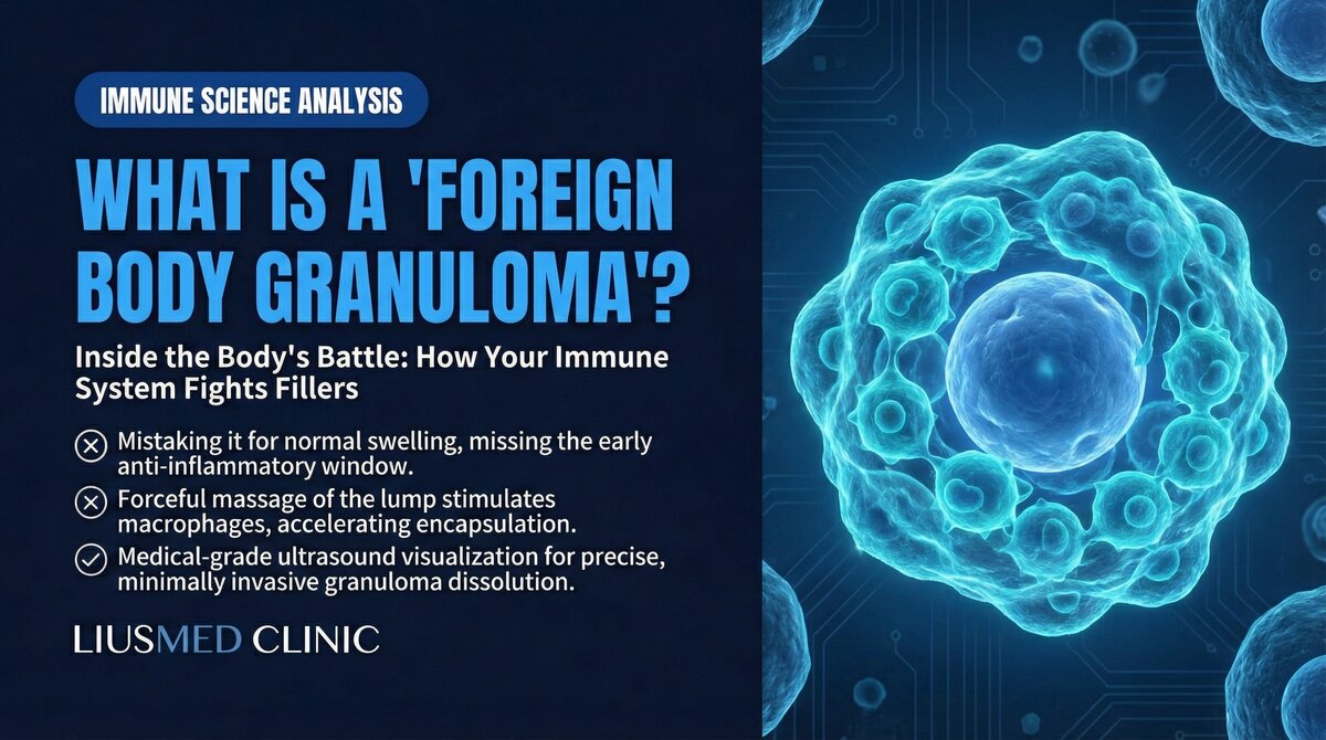
When Your Immune System Decides "This Doesn't Belong Here"
Months or even years after a filler injection, a hard, red, persistently swollen nodule appears at the injection site. It is not an infection, nor is it simple filler displacement. This is your immune system launching a full-scale war against a foreign material it cannot break down. In medical terminology, this reaction is called a foreign body granuloma.
Many patients panic when they first experience a granuloma: "Is it a tumor?" or "Was there something wrong with the filler?" In reality, a granuloma is a normal immune defense mechanism — but in the context of dermal fillers, this defense becomes an endless war of attrition with no winner.
Key Insight: A foreign body granuloma is neither an allergic reaction nor an infection. It is your immune system's chronic siege strategy — an attempt to "digest" a foreign substance it can never break down. It is a war your body can never win.
How the Immune System Recognizes Foreign Materials
The First Line of Innate Immunity
When any foreign substance enters tissue, the immune response follows a precise, stepwise escalation:
-
Damage signal release: The injection itself causes micro-tissue injury. Damaged cells release Danger-Associated Molecular Patterns (DAMPs), alerting nearby immune cells.
-
Neutrophil first responders: Within hours, neutrophils arrive at the injection site and attempt to engulf and destroy the foreign material. Against small invaders like bacteria, this is usually sufficient.
-
Macrophage takeover: When neutrophils cannot handle the material — filler particles are far larger than bacteria — monocytes are recruited from the bloodstream and differentiate into macrophages to take over the cleanup.
-
The "can't digest it" dilemma: Macrophages attempt to phagocytose filler particles, but these synthetic materials cannot be broken down by lysosomal enzymes. The macrophage is stuck — it has captured the enemy but cannot destroy it.
From Acute Inflammation to Chronic Warfare
This "can't digest it" dilemma is the turning point of the entire story.
| Immune Response Phase | Timeline | Primary Cells | Clinical Presentation |
|---|---|---|---|
| Acute inflammation | Hours to days | Neutrophils | Mild redness, normal post-injection reaction |
| Chronic inflammation onset | Days to weeks | Macrophages | Persistent swelling, pressure sensitivity |
| Giant cell formation | Weeks to months | Multinucleated giant cells | Palpable hard nodule |
| Granuloma maturation | Months to years | Organized granuloma | Firm mass, skin discoloration |
| Fibrotic encapsulation | Years | Fibroblasts | Permanent hard lump, tissue distortion |
The Molecular Mechanisms of Granuloma Formation
Giant Cell Fusion: A Desperate Escalation Strategy
When individual macrophages cannot engulf larger filler particles, the immune system activates an unusual strategy: multiple macrophages fuse into a single, massive multinucleated giant cell, also known as a Foreign Body Giant Cell (FBGC).
This fusion process is regulated by multiple cytokines:
- IL-4 and IL-13: Secreted by T-helper cells, these promote macrophage fusion
- RANKL: Promotes formation of osteoclast-like giant cells
- MCP-1 (CCL2): Continuously recruits more monocytes from the bloodstream
- TNF-alpha: Maintains the chronic inflammatory environment
Key Insight: The foreign body giant cell is the immune system's "ultimate weapon" — when regular troops cannot handle the enemy, soldiers fuse into a giant. But even a giant cannot digest synthetic filler materials.
The Organizational Structure of a Granuloma
A mature foreign body granuloma has a highly organized structure, much like a fortress built around an enemy:
- Core zone: Filler particles tightly surrounded by foreign body giant cells and macrophages
- Lymphocyte ring: T-cells and B-cells forming an outer defense perimeter, continuously secreting inflammatory mediators
- Fibroblast layer: The outermost layer, depositing collagen to form a fibrous capsule — effectively building a wall around the enemy
- Neovascularization: New blood vessels supplying the "battlefield" with a continuous stream of immune cells and nutrients
This explains why granulomas feel hard and well-defined to the touch — they are not a chaotic mass of tissue, but a carefully constructed immune fortification.
Granuloma Risk Differences Among Filler Types
Chemical composition, particle size and surface properties all affect how strongly the immune system reacts. One thing to be clear about first: there is no table that ranks granuloma rates across filler types. Every study enrolls different people, defines nodules differently and follows patients for different lengths of time, so lining those numbers up looks tidy while comparing things that are not comparable.
| Filler Type | Why the Immune System Targets It | What to Watch for Clinically |
|---|---|---|
| Hyaluronic acid (HA) | Cross-linker residues or protein contamination | Enzyme can dissolve it, but may not reach material inside an established capsule |
| Poly-L-lactic acid (PLLA/Sculptra) | Particulate form readily triggers giant cell response | Reconstitution method and injection technique both affect nodule risk |
| Polycaprolactone (PCL/Ellanse) | Large microsphere surface area, slow degradation | Material stays in tissue for a long time, so assess lumps with that in mind |
| Calcium hydroxylapatite (CaHA/Radiesse) | Uneven microsphere degradation | Ultrasound appearance differs from other lump types |
| Silicone / permanent fillers | Permanent foreign body that cannot be metabolized | Reactions can appear many years after injection |
| Autologous fat | Oil cysts forming after fat necrosis | Separate it from infection and cysts before deciding on treatment |
Every figure you can look up is tied to the conditions of its own study. A systematic review of collagen-stimulating fillers pooled 40 reports covering 117 patients who had already developed foreign body granulomas. The case mix was PMMA (Polymethyl Methacrylate, a non-absorbable microsphere that stays in tissue long term) 41/117 (35.04%), PLLA 36/117 (30.77%), CaHA 32/117 (27.35%), PCL 5/117 (4.27%) and dextran-based filler 3/117 (2.56%); nodules were the most common presentation (82.91%), the interval from injection to detection ranged from one week to 15 years (mean 20.18 months), and the perioral region was the most frequently reported site (Wang et al., Journal of Cosmetic Dermatology, 2025, PMID 41132036). That is the make-up of patients who already had a problem, and it cannot be turned around into an incidence rate for someone considering injection.
For HA, a retrospective chart review of Voluma recorded 23 patients with delayed-onset nodules across 4,702 treatments over 68 months, with a median of four months from injection to reaction (Beleznay et al., Dermatologic Surgery, 2015, PMID 26166260). The paper reports that as 0.5%, but the numerator counts patients while the denominator counts treatments, so the ratio is descriptive rather than a personal risk figure. PLLA has two cosmetic studies run under different conditions: 4 nodules among 1,002 patients (0.4%; Palm et al., Journal of Drugs in Dermatology, 2021, PMID 33400413), and 4.7% among 106 patients followed for two years (Schierle and Casas, Aesthetic Surgery Journal, 2011, PMID 21239677). For PCL, CaHA, silicone and autologous fat there are no comparable cosmetic-population rates to cite; PCL accounted for only five cases in the review above. Even supposedly "absorbable" fillers carry a non-zero granuloma risk. Cross-linker residues in HA (Hyaluronic Acid — sugar molecule naturally in skin, holds water), particle aggregation in PLLA (Poly-L-Lactic Acid (Sculptra) — particle injection stimulating collagen), and the surface properties of PCL (Polycaprolactone (Ellansé) — longer-lasting collagen stimulator) microspheres can all become targets that the immune system locks onto.
Differentiating Granulomas from Other Filler Lumps
When patients feel a hard lump at an injection site, there can be multiple causes. Accurate differential diagnosis is essential for choosing the right treatment approach.
Common Types of Filler Lumps
- Filler material nodules: Visible lumps from filler aggregation or superficial placement, usually without inflammatory signs
- Encapsulation: Fibrous tissue wrapping around filler, forming a smooth, mobile hard lump
- Biofilm-related swelling: Recurrent swelling caused by bacterial biofilm, often with intermittent flare-up patterns
- RE2O human-dermal-matrix skin-booster nodules: Papules or a deep consolidated lump from the Korean particulated human-ADM skin booster; hyaluronidase dissolves only the carrier half, so it needs ultrasound typing then pinhole removal
- Foreign body granuloma: Immune-mediated chronic inflammatory nodule, typically firm with poorly defined borders merging into surrounding tissue
The Diagnostic Value of High-Resolution Ultrasound
Among these different causes, high-resolution ultrasound is the preferred non-invasive diagnostic tool. Ultrasound can reveal:
- The exact location, depth, and distribution of filler material
- The degree of surrounding tissue inflammation and blood flow changes
- Whether capsular structures or liquefactive necrosis are present
- Aggregation patterns of filler particles
This "see before you treat" approach is the key to avoiding blind treatments that cause additional harm. To learn more about ultrasound in filler diagnosis, see the filler repair evaluation process.
Why Traditional Medical Treatments Have Limited Effectiveness
The Temporary Nature of Steroid Injections
Local steroid injection (such as Triamcinolone) is the most common first-line treatment for granulomas. Steroids work by suppressing inflammatory responses and immune cell activity to reduce granuloma volume.
The problem is that steroids only temporarily suppress the immune response without removing the root cause — the filler material itself. Once steroid effects wear off, the immune system re-engages, and the granuloma often recurs.
Worse, repeated steroid injections can cause serious side effects:
- Local fat atrophy (skin depression)
- Skin thinning and telangiectasia
- Pigmentation abnormalities
- Impaired tissue repair capacity
The Limitations of 5-FU
5-Fluorouracil (5-FU), as an antimetabolite, can inhibit fibroblast proliferation and has some effect on fibrosis-dominant granulomas. However, it also cannot eliminate existing filler material, and in treating collagen stimulator nodules, its results are often disappointing.
The Misuse of Antibiotics
When granulomas are misdiagnosed as infections, patients may receive multiple rounds of antibiotics. Since granulomas are fundamentally immune reactions rather than infections, antibiotics are not only ineffective but can cause side effects such as gut dysbiosis and delay appropriate treatment.
Key Insight: All medical treatments "manage symptoms" rather than "solve the problem." As long as the filler material triggering the immune response remains in the tissue, the granuloma retains the potential to recur. The logic of a cure is simple — remove the foreign body.
Why Physical Removal Is the Definitive Solution: An Immunological Perspective
Immunology teaches us a simple truth: a foreign body granuloma exists because of the foreign body. Remove the foreign body, and you remove the target that the immune system is continuously attacking.
However, removing filler is not as straightforward as it might seem. Traditional methods include:
- Hyaluronidase dissolution: Only effective for HA, and even then, the enzyme often cannot penetrate the capsule to reach the filler core in encapsulated cases (see Encapsulation: Why Dissolvers Fail)
- Blind squeezing or aspiration: Operating without seeing the exact filler location risks damaging normal tissue
- Surgical excision: While effective at removing filler, the resulting scars and tissue damage may be worse than the original problem
Ultrasound-guided minimally invasive extraction offers a more precise option. Under real-time high-resolution ultrasound guidance, the physician can pinpoint every filler deposit's location and depth, extracting material through a tiny pinhole incision while maximizing preservation of surrounding normal tissue.
This "see before you treat" principle not only improves clearance rates but also dramatically reduces collateral tissue damage. Learn more about the filler lump extraction technique.
Prevention and Early Recognition of Granulomas
Strategies to Reduce Granuloma Risk
While granuloma formation cannot be completely prevented, the following measures can reduce risk:
- Choose quality filler products: Products with low impurity levels and stable manufacturing are less likely to trigger immune reactions
- Avoid excessive injection volumes: The more filler material in the tissue, the greater the burden on the immune system
- Strict aseptic technique: Reducing bacterial contamination prevents biofilm formation and subsequent biofilm-related swelling
- Carefully assess injection sites: Areas with pre-existing foreign material carry higher risk with repeat injection
When Should You Seek Medical Attention?
If you experience any of the following after filler injection, seek professional evaluation promptly:
- A gradually enlarging firm nodule at the injection site
- A nodule accompanied by persistent redness or increased skin temperature
- New lumps appearing months to years after injection
- Sudden changes in a previously stable injection area
Early detection and accurate diagnosis can prevent unnecessary medication cycles and allow intervention before tissue damage becomes extensive. If you are considering filler repair, please contact us for an evaluation.
Common questions
Is a granuloma a tumor? Could it be cancer?
No. When people feel a hard lump, their first fear is often a tumor, but a foreign body granuloma is really your immune system reacting to filler it cannot break down — an inflammatory response, not cancer. That said, any new lump at an injection site is still worth having a doctor look at, just to be sure.
Why does the lump show up months or even years after the filler injection?
Because a granuloma builds up slowly over time rather than right after the injection. Immune cells keep trying to clear the filler and cannot, and eventually they wall it off into a nodule. The timing varies with the material too — some show up within a few months, others take a year or two, or even longer.
Can a steroid injection make the granuloma go away?
A steroid can calm the inflammation and shrink the nodule for a while, but it does not remove the filler that is driving the reaction. Once the steroid wears off, the immune system tends to start up again and the nodule often comes back. Repeated steroid shots can also thin the skin or leave a dent, so they manage the symptom rather than address the cause.
Do only hyaluronic acid fillers cause granulomas?
No. Even fillers described as absorbable carry a risk that is not zero. Hyaluronic acid, collagen stimulators, calcium hydroxylapatite, silicone, and even your own fat each carry their own risk, just at different rates. The immune system reacts to any foreign material it cannot break down, not just one type.
How can I tell whether my lump is a granuloma or something else?
Feeling it by hand is not reliable, because a filler nodule, a capsule, and biofilm swelling can all feel similar to a granuloma. In the clinic we use high-resolution ultrasound to see the filler's position and depth and how inflamed the area is, then work out which one it is and how to handle it. Looking first is safer than treating blind.
Understanding the Immune Mechanism Is the Foundation for Sound Treatment Decisions
Foreign body granulomas are not medical accidents — they are your immune system activating an ancient and effective defense strategy against a substance it cannot digest. Understanding this mechanism helps patients make more rational treatment decisions:
- Medication can temporarily control symptoms but cannot eliminate the cause
- Repeated pharmacotherapy may cause additional tissue damage
- Physical removal of the foreign body is the only way to break the immune response cycle
- Precise image guidance makes the removal process safer and more effective
Your immune system is faithfully doing its job to protect you. The solution is not to suppress it, but to remove the reason it keeps fighting. Learn more about our filler repair services.

Specialties
Credentials
- Kaohsiung Medical University, School of Medicine
- Attending Physician, Dermatology, Kaohsiung Chang Gung Memorial Hospital
- Attending Physician, Aesthetic Center, Kaohsiung Chang Gung Memorial Hospital
- Visiting Physician, Dermatology, Xiamen Chang Gung Hospital
- Visiting Physician, Aesthetic Center, Xiamen Chang Gung Hospital
"For every surgery, I strive to achieve a good outcome through a small incision and refined technique. Minimally invasive surgery is not just a technique — it's a commitment of respect to every patient."
Recovery after any procedure needs peer support too


Want to learn more?
Schedule a consultation for professional evaluation and advice