How Do We Confirm Complete Apocrine Gland Removal? Intraoperative Visualization and Clinical Follow-Up

"Did you get all of it?" That's one of the most common questions after axillary odor surgery, and honestly one of the harder ones to answer in a single sentence.
Apocrine glands, the large sweat glands responsible for body odor, are scattered just beneath the dermis in the axilla. They're completely invisible from the skin surface, and their distribution, depth, and density vary significantly between individuals. So "complete removal" isn't a fixed threshold. It depends on what your apocrine anatomy looks like, how the surgeon confirms what's been cleared, and what happens during follow-up.
Here's how we approach that confirmation at Liusmed Clinic.
Why This Matters: The Glands You Can't See
Apocrine glands sit just below the dermis, in the superficial fatty layer of the axilla. A closed wound after surgery doesn't tell you whether the glandular tissue underneath has been thoroughly addressed. Traditional blind curettage (where the surgeon scrapes by feel alone) has a structural limitation: there's no real-time visual feedback during the procedure.
That's not a criticism of technique. It's a limitation of the method itself. Even a careful surgeon working without visualization can't confirm in real time whether the excision plane is reaching the target depth, or whether the margins are covered.
Residual apocrine tissue is the primary mechanism behind odor recurrence after surgery. For a deeper explanation of how residual glands cause symptoms to return, see Why Axillary Odor Surgery Sometimes Fails to Last.
Intraoperative Confirmation Layer One: Ultrasound Mapping Before the First Incision
At Liusmed, the surgical workflow begins with preoperative ultrasound scanning.
Ultrasound clearly shows the depth, distribution, and thickness of the apocrine layer, before the incision is made. The surgeon enters the procedure knowing exactly where this patient's glands are concentrated, how deep they sit, and where the margins extend. In traditional surgery, none of this information exists. The surgeon estimates by feel as they go.
Ultrasound guidance means the excision follows the target tissue rather than relying on probability. For patients with an unusually thin glandular layer, or glands extending toward the peripheral margins, this makes a meaningful difference in coverage.
Key point: The value of ultrasound guidance isn't just precision. It's that the surgeon has real-time information about what they're clearing. Without imaging, what's missing is visual feedback, not skill.
Intraoperative Confirmation Layer Two: Direct Inspection of the Excision Plane
Once ultrasound has confirmed the direction, the rotary cutter enters through a small incision (typically less than 20% of the lesion's diameter), and the surgeon can directly inspect the excision plane as it develops.
This direct view is what distinguishes minimally invasive technique from blind curettage. The incision is small, but the confirmation is active and ongoing: each zone is inspected before moving to the next, allowing systematic coverage of both the central area and the margins.
For a detailed explanation of rotary cutter technique and how it achieves complete apocrine gland clearance, see Rotary Cutter Axillary Surgery: How It Works.
Comparison: Clearance Confirmation in Traditional vs. Ultrasound-Guided Surgery
| Confirmation step | Traditional blind curettage | Ultrasound-guided rotary cutter |
|---|---|---|
| Preoperative gland depth mapping | None | Ultrasound localization |
| Intraoperative range confirmation | Tactile estimate | Real-time imaging guidance |
| Direct excision plane inspection | Not possible (blind) | Yes, surgeon inspects directly |
| Peripheral margin assessment | Subjective judgment | Ultrasound re-scan possible |
| Compensation for anatomy variation | Difficult | Adjusted per individual anatomy |
| Post-excision verification | Wait for clinical follow-up | Intraoperative ultrasound check available |
Postoperative Confirmation: What Clinical Follow-Up Is Monitoring
Surgery isn't the end of the confirmation process. Follow-up has several layers:
First, wound healing. Compression dressing and proper skin flap approximation are fundamental. Detailed care guidance is in the Postoperative Wound Care Guide.
Second, once healing stabilizes (typically at the 1- to 2-month mark), the actual effect on odor becomes assessable. Early swelling affects tissue behavior, so evaluating outcomes before the 4- to 6-week window isn't reliable.
Third, if noticeable odor persists beyond the 2- to 3-month mark, ultrasound can reassess whether residual glandular tissue remains. When residual tissue is identified, supplementary clearance may be appropriate in some cases.
Key point: What follow-up actually looks for is whether persistently active residual glands remain. On long-term clinical follow-up, odor returning after a thorough clearance has been uncommon. Gland activity still varies between individuals, so results vary too.
Why Individual Anatomy Shapes the Outcome
The number, density, and activity level of apocrine glands is largely genetic and varies considerably between individuals. Some patients have glands concentrated in the central axillary zone, which is straightforward to cover systematically. Others have glands extending into peripheral areas that require more careful attention to margins.
This is why preoperative ultrasound evaluation matters. Not every patient's apocrine anatomy is the same. Knowing the distribution before the incision is made, rather than adjusting intraoperatively without imaging, produces more predictable results.
If your outcome after surgery feels lower than expected, the right first step is a follow-up assessment, not guesswork. Ultrasound can distinguish between residual glandular tissue, individual gland activity levels, and other factors, which determines what the appropriate next step is.
If you have questions about what to expect, feel free to contact the clinic.
Common Questions
How long after surgery can you reliably assess the outcome?
One to two months after surgery, once swelling has settled and tissue has stabilized, is the appropriate window for outcome assessment. Early odor changes during the healing phase aren't reliable indicators. Give it time before drawing conclusions.
If the outcome isn't satisfactory, can supplementary treatment be done?
Yes, but only after follow-up ultrasound evaluation to determine whether residual tissue is the cause. If residual apocrine glands are identified, supplementary clearance is an option in some cases. If the issue is individual gland activity rather than incomplete removal, a different approach may be needed. Starting supplementary treatment without a clear diagnosis can mean addressing the wrong problem.
Is ultrasound-guided surgery suitable for everyone?
Patients who are appropriate candidates for minimally invasive axillary odor surgery can generally benefit from ultrasound guidance. But suitability, including which specific approach makes sense, requires an in-clinic evaluation. Individual anatomy and circumstances vary.
Can the odor come back after surgery?
The main mechanism behind odor returning is residual apocrine tissue, which is exactly why clearance is confirmed during surgery and monitored afterwards. On long-term clinical follow-up, odor returning after a thorough clearance has been uncommon, though individual gland activity differs, so results vary. If noticeable odor persists 2 to 3 months after surgery, an ultrasound check can tell whether residual tissue is the cause.
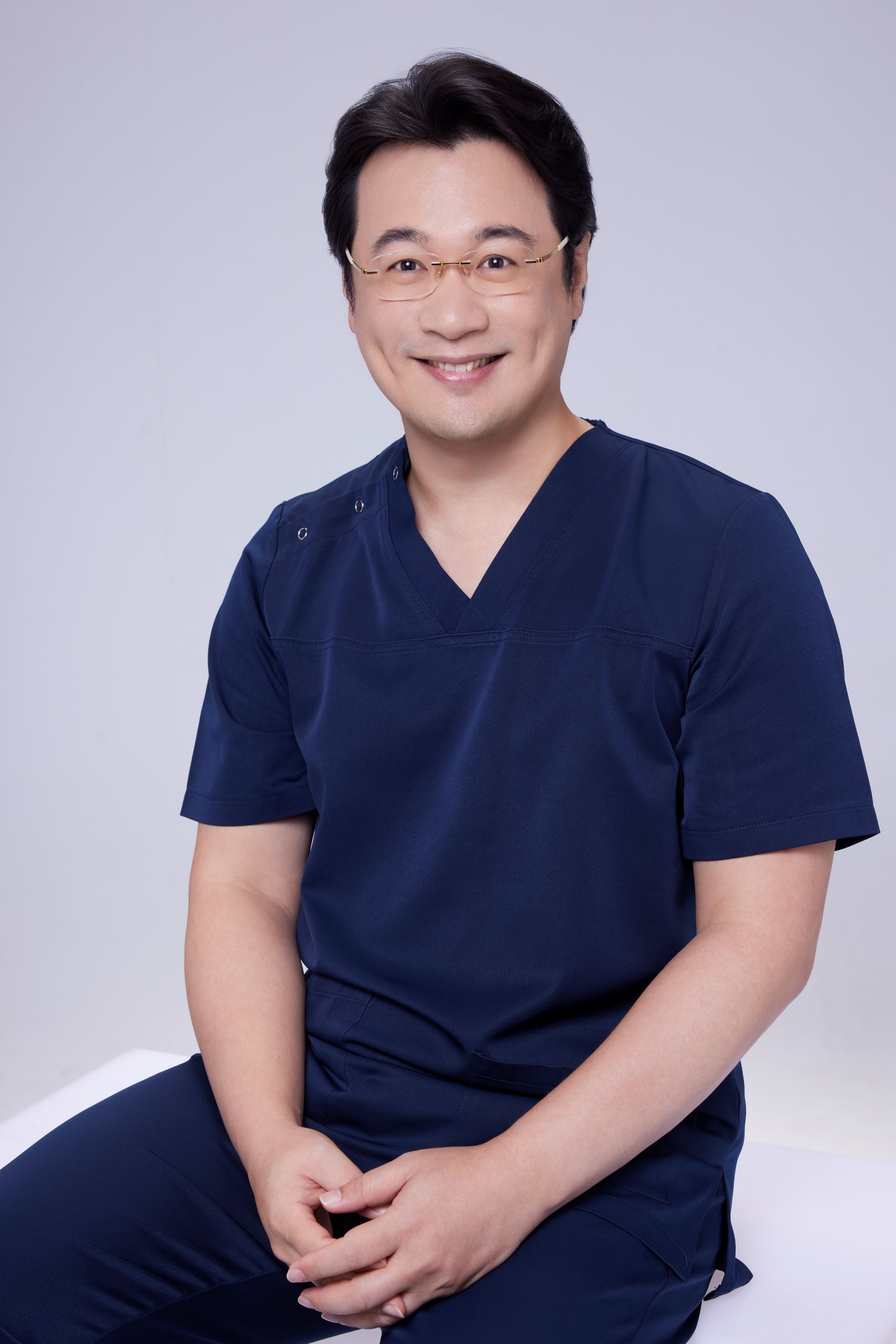
Medically reviewed by Dr. Ta-Ju Liu. Written in accordance with Taiwan's Medical Care Act. This article is educational and does not constitute personal medical advice. Please consult a physician for individualized treatment planning.
Specialties
Credentials
- Kaohsiung Medical University, School of Medicine
- Attending Physician, Dermatology, Kaohsiung Chang Gung Memorial Hospital
- Attending Physician, Aesthetic Center, Kaohsiung Chang Gung Memorial Hospital
- Visiting Physician, Dermatology, Xiamen Chang Gung Hospital
- Visiting Physician, Aesthetic Center, Xiamen Chang Gung Hospital
"For every surgery, I strive to achieve a good outcome through a small incision and refined technique. Minimally invasive surgery is not just a technique — it's a commitment of respect to every patient."
Want to learn more?
Schedule a consultation for professional evaluation and advice