Why Does a Cyst Recur After Removal? Incomplete Excision
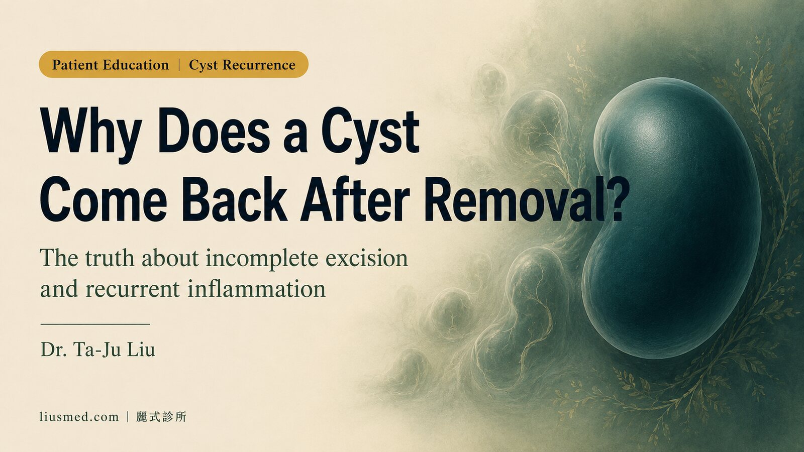
It's a scenario we see often in clinic: a patient comes in with visible frustration and a familiar lump in a familiar location. "I had this removed a few years ago. Why is it back? Is this just how my body works?"
The answer is almost always the same: the cyst came back because the cyst wall was not completely removed.
This is not a condemnation of the previous procedure — complete wall excision can be genuinely difficult, especially after inflammation has fused the capsule to surrounding tissue. But understanding why the cyst recurred gives you the clearest path forward.
The Core Biology: A Cyst Is a Self-Filling Sac
To understand recurrence, you first need to understand what an epidermal cyst (epidermal cyst, also called sebaceous cyst in common usage) actually is.
An epidermal cyst is a closed sac lined with squamous epithelium — the same cell type as your outer skin. That lining continuously sheds keratin (cornified cells), exactly as normal skin does. But because the sac has no functional opening, the shed material cannot escape; it accumulates inside, causing the cyst to grow.
The key insight: the contents of a cyst are a byproduct of the cyst wall. They are not the problem itself.
If you remove the contents but leave the epithelial lining, that lining keeps producing keratin. The cyst refills. Recurrence is not a question of if, but when.
Key point: Clearing the cyst's contents while leaving the wall is like repeatedly emptying a leaking pipe by mopping the floor — helpful in the moment, but the water keeps coming.
Three Common Paths to Incomplete Removal
1. Self-squeezing
Squeezing a cyst may express some of the white, cheese-like keratin through the punctum (the small central pore visible on some cysts). But the cyst wall remains untouched beneath the skin and continues producing keratin.
What's more, the mechanical pressure of squeezing can rupture the cyst wall at depth, triggering a foreign-body inflammatory reaction in the surrounding tissue. This creates dense adhesions between the cyst wall and the dermis, making the next excision harder.
2. Incision and drainage (I&D)
When a cyst becomes acutely infected and inflamed, incision and drainage (I&D) is sometimes performed to decompress the abscess and relieve pain. This is a legitimate acute intervention — but its purpose is infection control, not cure.
After drainage, the cyst wall remains. As inflammation resolves, the cyst almost always reforms.
Key point: The correct sequence for an inflamed cyst is: I&D (or antibiotics) to control acute infection → wait for full resolution → schedule complete excision. Attempting curative excision during active inflammation dramatically increases the risk of capsule rupture and incomplete removal.
3. Incomplete surgical excision
Even when surgery is performed, recurrence can occur if any portion of the cyst wall is left behind — for example, if the capsule ruptures intraoperatively and fragments remain in the wound, or if the surgical exposure is insufficient for the surgeon to clearly visualize and dissect the entire wall.
Why is complete capsule excision difficult in certain cases?
- Post-inflammatory adhesion: After one or more episodes of inflammation, the capsule becomes densely adherent to surrounding tissue. The clean dissection plane between cyst wall and dermis disappears.
- Difficult anatomical location: Sites near important structures (facial nerve, vessels) require conservative dissection, limiting maneuverability.
- Intraoperative rupture: Once the capsule tears, keratin spills into the wound, obscuring the residual wall fragments.
- Undersized incision: A very small incision limits visualization and makes it hard to confirm complete excision.
How Repeated Inflammation Makes Each Subsequent Removal Harder
Each time a cyst inflames — from self-rupture, squeezing, or infection — the inflammatory process generates scar tissue between the cyst wall and the surrounding dermis and fat.
Before any inflammation, there is a natural tissue plane between the cyst capsule and surrounding tissue. An experienced surgeon can dissect along this plane and deliver the cyst intact, like shelling a nut.
After inflammation, scar tissue obliterates this plane. The capsule and surrounding tissue become a single fused mass. Complete excision becomes technically more demanding, and the risk of intraoperative rupture increases with each prior episode of inflammation.
Key point: This is why treating a cyst before it inflames — when the natural dissection plane is intact — offers the best chance of complete excision and lowest recurrence risk.
Complete vs. Incomplete Removal: A Comparison
| Squeezing / Drainage | Incomplete Excision | Complete Excision | |
|---|---|---|---|
| What is removed | Contents only | Contents + partial wall | Contents + complete wall |
| Cyst wall remains | Yes | Partially | No |
| Recurrence | Near-certain | High (depends on residual) | Very low |
| Effect on future surgery | Increases difficulty | May increase difficulty | N/A |
| Appropriate timing | Acute emergency only | Not preferred | Stable (non-inflamed) cyst |
What "Complete Excision" Requires Technically
The surgical goal is to deliver the cyst as an intact structure, with the epithelial lining unruptured and entirely removed. This depends on:
- Adequate incision — sufficient exposure for the surgeon to visualize and dissect the full circumference of the capsule
- Pre-operative ultrasound — maps cyst depth, diameter, and relationship to adjacent structures; essential for planning, especially in recurrent or post-inflammatory cysts
- Dissection along the capsule wall — patient, layer-by-layer separation along the natural tissue plane, avoiding intraoperative rupture
- Confirming completeness — examining the excised specimen to verify the capsule is intact; if the wall was breached intraoperatively, any visible wall fragments must be retrieved
For cysts with prior inflammation or prior recurrence, ultrasound-guided (ultrasound-guided) pre-operative planning is especially important: it lets the surgeon see the exact dimensions of the cyst and the extent of adhesion before making any incision.
If Your Cyst Has Already Recurred: What Next?
Recurrence is treatable — repeat complete excision is effective, though technically more demanding than first-time removal.
Some guidance:
- Get a clear picture of the current state: Ultrasound can delineate the recurrent cyst's size, depth, and the extent of adhesion to surrounding tissue — providing the foundation for surgical planning.
- Treat active inflammation first: If the cyst is currently inflamed, the priority is resolution (antibiotics and/or drainage), not immediate excision. Operating during active inflammation dramatically increases the risk of incomplete removal.
- Share your history: Tell your surgeon how the cyst was treated before — drainage, how many excisions, any episodes of significant post-surgical inflammation. This helps risk stratification.
If your cyst has recurred once or multiple times, ultrasound evaluation before cyst excision is a critical first step. For select recurrent cysts, laser cyst removal may also be an option — suitability depends on individual anatomy and the degree of prior scarring. You are also welcome to explore the full range of skin tumor care at our clinic.
Common questions
My cyst grew back in the same spot after removal — is that just how my body is?
Almost never. The usual reason a cyst comes back is that the wall of the sac wasn't fully taken out the first time. As long as that lining is still under the skin making keratin, the sac slowly fills again and the lump returns.
Can I just squeeze the cyst out myself?
Squeezing might push out some of the contents, but the wall stays put under the skin and keeps working. Worse, pressing hard can rupture the wall deep down, set off inflammation and adhesions to the surrounding tissue, and make a proper excision later harder.
Can the cyst be excised while it's inflamed or infected?
Operating during an active flare-up isn't ideal. When it's inflamed the wall is stuck to the surrounding tissue, so cutting it out then carries more risk and leaves fragments behind more easily. Usually we settle the inflammation first with drainage or medication, then schedule the full excision once things are calm.
Is a cyst that has flared up several times harder to treat?
Yes. Each episode of inflammation lays down more scar tissue around the wall, blurring the clean plane the surgeon normally works along, so the wall tears more easily during surgery. That's why treating it in a stable, non-inflamed period gives a better chance of taking the whole wall out.
My cyst has already recurred — can it still be dealt with, and what happens before surgery?
Yes. A repeat complete excision works well, it's just technically more demanding than the first time. Beforehand an ultrasound usually maps how big it is and how much it's stuck down; if it's inflamed right now that gets settled first, and it helps to tell your surgeon how it was treated before.
In Summary
Cyst recurrence is almost never a matter of "just the way your body is." It almost always has a specific structural cause: the cyst wall was not completely removed.
Understanding that cause allows you to have a clearer conversation with your surgeon and choose a treatment path that addresses the root, not just the symptom.
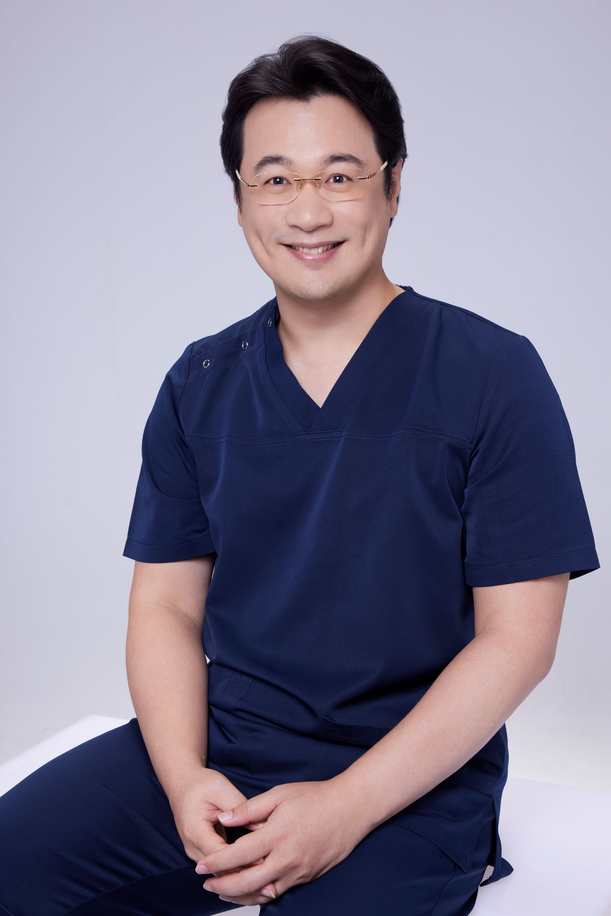
If you have questions about a recurring cyst or want a surgical evaluation, book a consultation. Dr. Ta-Ju Liu at LIUSMED Clinic will assess your individual situation and recommend the most appropriate approach.
Further reading
Specialties
Credentials
- Kaohsiung Medical University, School of Medicine
- Attending Physician, Dermatology, Kaohsiung Chang Gung Memorial Hospital
- Attending Physician, Aesthetic Center, Kaohsiung Chang Gung Memorial Hospital
- Visiting Physician, Dermatology, Xiamen Chang Gung Hospital
- Visiting Physician, Aesthetic Center, Xiamen Chang Gung Hospital
"For every surgery, I strive to achieve a good outcome through a small incision and refined technique. Minimally invasive surgery is not just a technique — it's a commitment of respect to every patient."
Want to learn more?
Schedule a consultation for professional evaluation and advice