Layer-by-Layer Extraction Strategy When Filler Is Distributed Across Multiple Tissue Planes
Why Does Filler Spread Across Multiple Layers?
Ideally, filler should remain at its target injection plane. In practice, however, filler may distribute across different tissue depths for multiple reasons, creating complex multi-layer patterns.
Causes of Multi-Layer Distribution
| Cause | Mechanism | Common Scenario |
|---|---|---|
| Imprecise injection depth | Different sessions entering different depths | Repeated injections to the same area |
| Filler migration | Gravity, muscle movement, external pressure | Nose, forehead, zygomatic region |
| Post-dissolution redistribution | Residual migrates after partial dissolving | After failed hyaluronidase treatment |
| Different injectors | Different physicians using different techniques and planes | Treatment at multiple clinics |
| Material properties | Liquid fillers naturally diffuse | Liquid silicone, dilute HA |
For more on migration mechanisms, see How Fillers Migrate.
Key Insight: Multi-layer filler distribution is one of the most challenging revision scenarios. Filler at different depths requires different extraction strategies, and a "one-size-fits-all" approach simply cannot address this complexity. Only ultrasound can precisely show filler position at each layer.
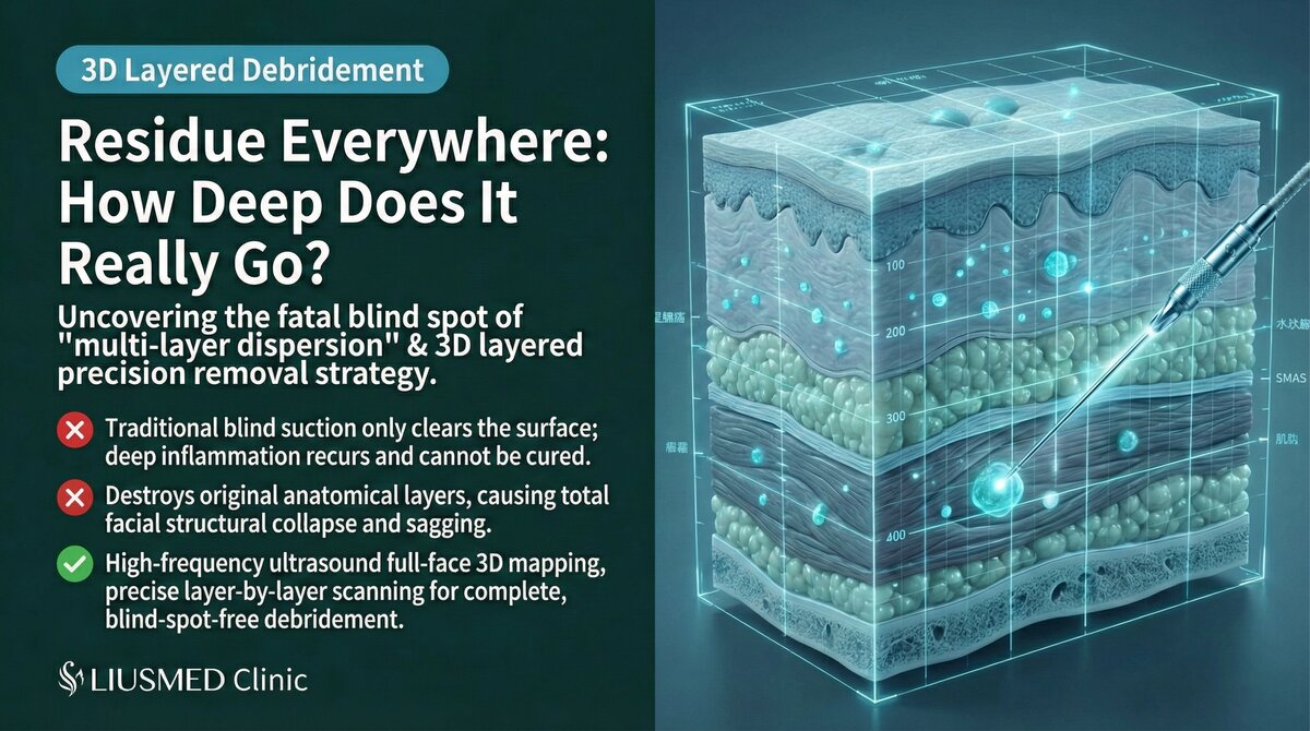
Facial Tissue Layer Architecture
Anatomical Layers to Understand
| Layer (Superficial to Deep) | Characteristics | Common Filler Location |
|---|---|---|
| Epidermis/Dermis | Shallowest, directly affects appearance | Superficial HA (tear trough, fine lines) |
| Superficial subcutaneous fat | Primary filler target plane | Most HA, Radiesse |
| SMAS (Superficial muscular aponeurotic system) | Important structural layer | Some deep fillers, migrated material |
| Deep subcutaneous fat | Deeper filler target | Sculptra, deep injections |
| Muscle layer | Contains facial expression muscles | Migrated filler |
| Deep fascia/Periosteum | Deepest layer | Supraperiosteal injections, deep migration |
How Ultrasound Identifies Filler at Each Layer
Ultrasound clearly displays interfaces between different layers and precisely localizes which layer contains filler:
| Identification Element | Ultrasound Presentation |
|---|---|
| Filler depth | Measures distance from skin surface to filler |
| Tissue plane | Determined by surrounding tissue characteristics |
| Relationship between layers | Simultaneously displays complete multi-layer distribution |
| Filler properties at each layer | Different layers may contain different filler materials |
Layer-by-Layer Extraction Strategy
Fundamental Principle: Superficial to Deep
Layer-by-layer extraction follows a superficial-to-deep principle:
- Superficial first: Removing superficial filler creates space for deeper operations
- Progressive image clarity: Removing superficial interference improves deep-layer imaging
- Graduated risk management: Deeper operations carry higher risk, addressed last
- Ongoing assessment: Evaluate necessity of going deeper after each layer
Extraction Techniques by Layer
| Layer | Extraction Method | Special Considerations |
|---|---|---|
| Dermal/Superficial subcutaneous | Micro-pinhole aspiration or curettage | Avoid skin damage, watch for Tyndall effect |
| Subcutaneous fat layer | Standard ultrasound-guided extraction | Most common extraction plane |
| SMAS layer | Meticulous dissection then extraction | Watch for facial nerve branches |
| Intramuscular | Ultrasound-precise localization then separation | Protect expression muscle function |
| Supraperiosteal | Deep ultrasound-guided extraction | Watch for deep vessels, limited operating space |
Technical Challenges of Layered Extraction
Key Challenges
| Challenge | Explanation | Solution |
|---|---|---|
| Inter-layer interference | Superficial filler may obscure deep-layer imaging | Extract superficial layer first |
| Blurred boundaries | Filler may cross layer boundaries | Dynamic ultrasound tracking |
| Mixed materials | Each layer may contain different materials | Adjust method per material type |
| Deep operation risk | More critical structures at depth | Continuous ultrasound neurovascular monitoring |
| Limited operating space | Micro-incision limits deep reach | Multi-angle entry or multiple ports |
Key Insight: Multi-layer filler extraction is not a "single surgery" but a systematically planned engineering project. Each layer requires a customized extraction strategy based on its depth, material, and location.
Post-Operative Management and Follow-Up
Post-Operative Features of Layered Extraction
Compared to single-layer extraction, layered extraction requires closer follow-up:
| Follow-Up Item | Timing | Purpose |
|---|---|---|
| Post-op ultrasound | 1–2 weeks | Confirm clearance status at each layer |
| Swelling monitoring | Ongoing 1–2 weeks | Multi-layer operations may cause more swelling |
| Functional assessment | 2–4 weeks | Confirm deep operations haven't affected function |
| Long-term follow-up | 1–3 months | Assess tissue remodeling and final results |
Conclusion: Precise Layer Identification Is the Key to Success
Multi-layer filler revision tests not only surgical technique but also the physician's deep understanding of facial anatomy and professional ultrasound image interpretation. Every successful multi-layer revision begins with an accurate "three-dimensional filler distribution map."
If you know or suspect your filler is distributed across multiple layers, a complete ultrasound assessment to understand the exact distribution is recommended before formulating a treatment plan. Contact Liusmed Clinic to arrange a consultation.
Related reading: Filler Lump Extraction Technique, Ultrasound-Guided Pinhole Extraction Explained
Related Services

Specialties
Credentials
- Kaohsiung Medical University, School of Medicine
- Attending Physician, Dermatology, Kaohsiung Chang Gung Memorial Hospital
- Attending Physician, Aesthetic Center, Kaohsiung Chang Gung Memorial Hospital
- Visiting Physician, Dermatology, Xiamen Chang Gung Hospital
- Visiting Physician, Aesthetic Center, Xiamen Chang Gung Hospital
"For every surgery, I strive to achieve a good outcome through a small incision and refined technique. Minimally invasive surgery is not just a technique — it's a commitment of respect to every patient."
Recovery after any procedure needs peer support too
Want to learn more?
Schedule a consultation for professional evaluation and advice