How Nerve Hydrodissection Works: Freeing a Trapped Nerve Under Ultrasound Guidance
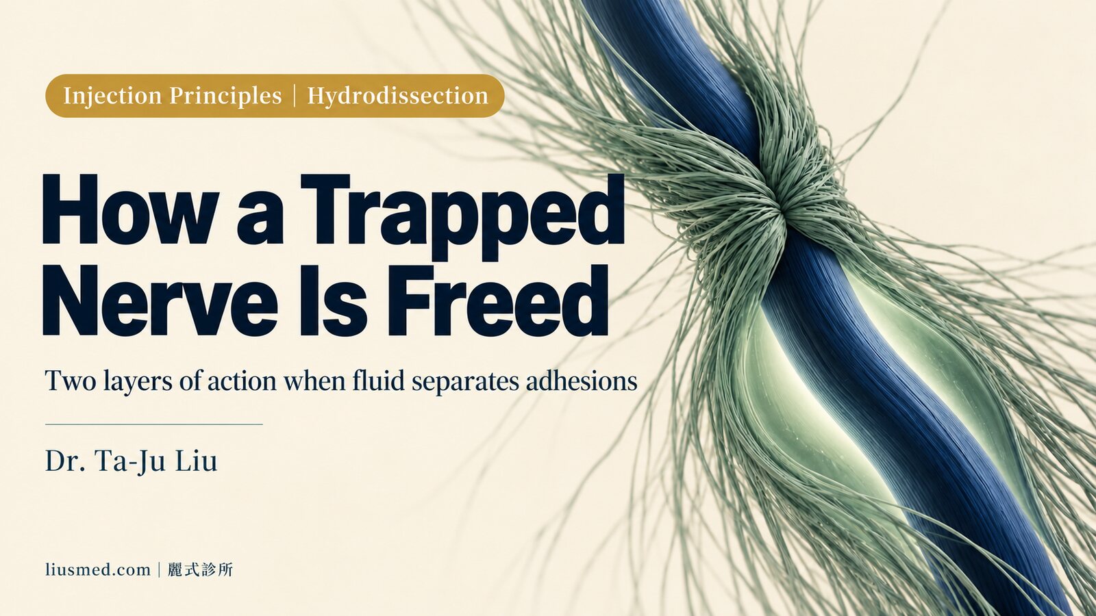
How Nerve Hydrodissection Works: Freeing a Trapped Nerve Under Ultrasound Guidance
I see this pattern often. Someone has had numbness in the hand for six months or more. They have taken medication, done rehab, worn a splint. It gets better for a while, then comes back. Imaging shows no severe compression, yet the numbness stays.
In cases like this the deciding factor is usually not how hard the nerve is being squeezed, but whether it can still glide. Nerve hydrodissection, an injection technique that uses fluid under imaging guidance to separate a nerve from the tissue around it, works on exactly that layer. Once you understand the mechanism you can judge more easily whether it suits your situation, and you will see why it is almost always done under ultrasound.
What happens when a nerve gets stuck
A peripheral nerve is not a pipe bolted into place. When your wrist bends, your fingers extend, your neck turns, the nerve has to glide between the surrounding fascia, tendons and connective tissue.
After long periods of postural load, repetitive use or an old injury, fibrotic adhesions form around the nerve. Once gliding is restricted, every movement tugs on the same segment. The nerve also has its own blood supply and sensory endings, the vasa nervorum and nervi nervorum. When those small structures are compressed, circulation and drainage suffer, metabolic waste accumulates locally, and what you feel is not only numbness but a dull ache you cannot quite point to.
Lam and colleagues set out this reasoning in their review, and note that the mechanical benefit of hydrodissection may relate to relieving compression of the nervi nervorum and vasa nervorum (PMID: 32801851, DOI 10.2147/JPR.S247208).
Key point: The numbness and pain often come from restricted gliding, not only from the degree of compression. That is also why someone whose imaging looks fine can still have obvious symptoms.
The mechanical layer: what the fluid actually does
Under ultrasound guidance, the needle tip is placed into the thin interface between the nerve and the tissue around it, and fluid is slowly injected. It spreads along the path of least resistance, opening up the adhesions, so that a gliding space reappears between the nerve and its neighbours.
One detail makes this clear. In the studies discussed below, even the control groups that received only normal saline showed significant improvement, which led the authors to conclude that part of the effect does come from purely mechanical decompression (PMID: 33746745, DOI 10.3389/fphar.2020.621150).
So the first layer of value here is giving the nerve its space back. It does not change the shape of bone, and it will not widen a tunnel that is structurally narrow to begin with. What it addresses is adhesion and gliding.
Choosing the fluid: why dextrose ranks well
The literature has compared several injectates: normal saline, local anaesthetic, corticosteroid, 5% dextrose in water, platelet-rich plasma, and in some studies hyaluronidase.
The systematic review mentioned above included ten studies, nine on carpal tunnel syndrome and one on ulnar neuropathy at the elbow. Dextrose and PRP showed consistently more favourable outcomes than control groups or corticosteroid, and no serious adverse events were reported (PMID: 33746745).
A 2025 network meta-analysis pooled nine studies and 458 patients with carpal tunnel syndrome and ranked the injectates using SUCRA. For Boston Carpal Tunnel Questionnaire function, 5% dextrose ranked first at 4, 12 and 24 weeks (SUCRA 99.9, 89.8 and 88.8). For symptoms, 5% dextrose ranked first at 4 weeks (99.9), while PRP ranked first at 12 and 24 weeks (95.7 and 93.9). On volume, a 5 cc injection ranked first for both domains (99.5). The authors also state that establishing precise protocols by disease severity needs further research (PMID: 39894044, DOI 10.3349/ymj.2024.0089).
Why dextrose might do a little more than saline alone is still being worked out. Proposed directions include downregulating TRPV1, an ion channel involved in pain signalling, and correcting a possible glucose shortfall around the nerve (PMID: 32801851). These remain hypotheses rather than settled mechanism.
Here is what that means for you. Each injectate matches a different goal and timeline, so choosing one is part of the assessment. A research ranking describes group averages, while your own response still varies with your tissue.
Key point: This procedure works on two layers at once, mechanical and pharmacological. Looking only at which fluid was injected misses half of it, and looking only at opening adhesions misses the other half.
Ultrasound guidance is the premise, not a bonus
A nerve is often only a few millimetres across, with an artery, tendons and other branches sitting right against it. Placing a needle tip into the interface between the nerve and its adhesions takes more than anatomical knowledge and surface landmarks.
Ultrasound provides three things at this step. First, you see the state of the nerve itself: an entrapped nerve is usually swollen compared with the same nerve on the other side, the cross-sectional area can be measured, and swollen fascicles are visible. Second, you see where the needle tip is the whole way in, instead of advancing on feel. Third, you see the fluid spreading in real time, so you know whether the adhesions are genuinely being opened or the fluid has gone into a different plane. Lam and colleagues list the in-plane approach, with needle and probe in the same plane, as the primary and safer method (PMID: 32801851).
Ultrasound-guided nerve repair work is built on this point. When you cannot see, you do not know what you have injected into, and that is a different order of risk from simply being imprecise.
How it sits beside steroid injection and surgery
These three approaches address different problems.
| Comparison | Nerve hydrodissection | Corticosteroid injection | Surgical decompression |
|---|---|---|---|
| Main goal | Separate adhesions, restore gliding | Suppress local inflammation and swelling | Release a structurally narrowed segment |
| Imaging guidance | Required, needle tip and fluid watched throughout | Recommended | Not applicable |
| Where it fits | Adhesion and restricted gliding predominate | Marked inflammation and swelling, faster relief needed | Clear structural narrowing, poor response to conservative care |
| Invasiveness | Injection | Injection | Surgery |
| Typical schedule | Staged by response, weeks apart | Number of doses limited | Single procedure |
In practice these mostly form a ladder, and which rung you start on depends on what is driving your symptoms. The full ladder for carpal tunnel syndrome is in the non-surgical treatment ladder for carpal tunnel syndrome. If you are still working out what your numbness and electric pain actually are, start with how nerve entrapment and sciatica work. A broader comparison of options sits in regenerative options for peripheral neuropathy.
What a session looks like
Assessment comes first. History plus ultrasound, to confirm the symptoms really do come from entrapment and adhesion of that nerve, and not from the cervical spine, the shoulder or a metabolic neuropathy wearing its clothes. Skip this step and accuracy later will not help, because you are treating the wrong target.
Then positioning and injection. The interface between nerve and adhesion is located, the needle tip travels along that layer, and fluid is delivered while you watch it push the adhesions apart. You will feel pressure during this; that is the fluid opening up space.
Afterwards the point is gliding. The nerve has to actually move through its new space for the adhesions to be less likely to reform, so light nerve gliding movements and ordinary daily activity are usually encouraged rather than complete rest. Heavy repetitive work and high-intensity training can wait a few days.
How quickly and how much you improve varies with individual tissue response. People whose symptoms have been running for a long time, with visible changes in the nerve, usually need staged treatment along with adjustments to posture and use.
Situations that need sorting out first
Some circumstances need handling or ruling out before an injection is scheduled:
- Infection at the injection site or systemically
- A clotting disorder, or current anticoagulant use
- Pregnancy
- Malignancy or a lump of unclear origin in the area
Diabetes on its own is not a reason to hold back: the usual 5 mL of 5% dextrose contains only about 0.25 g of glucose, so the effect on blood sugar is minor. Just let the doctor know beforehand.
Separately, please get these assessed before thinking about injections: obvious muscle weakness or wasting, change in bladder or bowel function, sudden loss of sensation after trauma, symptoms worsening rapidly over days, unexplained weight loss. Those point to other causes or to something that needs attention sooner. The full picture on indications and contraindications is in who is suitable for regenerative treatment.
Common questions
Is the injection going into the nerve itself?
No, it goes outside the nerve. The target is the interface between the nerve and the tissue around it, so the two can be separated. That is also why the needle tip is watched the whole way; entering the nerve is something to avoid.
Why dextrose rather than saline?
Saline works too, since the mechanical opening itself does something. But in studies comparing injectates, 5% dextrose generally ranked higher for both symptoms and function, possibly through effects on pain signalling channels. Which one fits depends on your situation and the goal of treatment.
Is one session enough?
It depends on how widespread the adhesions are and how long the symptoms have been running. Some people improve after one session and hold; others need staged treatment weeks apart, adjusted by how they respond. That expectation should be set at the assessment, not guessed at afterwards.
Is this the same as prolotherapy?
No. Prolotherapy targets the repair response in ligaments, tendons and joints, while nerve hydrodissection targets adhesion and gliding around a nerve. Sometimes both are used in one treatment plan for different problems.
Can it be done without ultrasound?
Technically some people do, but you lose the ability to tell where the needle tip is and where the fluid went, which is the core of the procedure. A few millimetres around a nerve is a different structure, and that is why I do not accept blind injection.
References
- Lam KHS, Hung CY, Chiang YP, et al. Ultrasound-Guided Nerve Hydrodissection for Pain Management: Rationale, Methods, Current Literature, and Theoretical Mechanisms. J Pain Res. 2020;13:1957-1968. PMID: 32801851. DOI: 10.2147/JPR.S247208
- Buntragulpoontawee M, Chang KV, Vitoonpong T, et al. The Effectiveness and Safety of Commonly Used Injectates for Ultrasound-Guided Hydrodissection Treatment of Peripheral Nerve Entrapment Syndromes: A Systematic Review. Front Pharmacol. 2020;11:621150. PMID: 33746745. DOI: 10.3389/fphar.2020.621150
- Lee K, Park JM, Yoon SY, et al. Ultrasound-Guided Nerve Hydrodissection for the Management of Carpal Tunnel Syndrome: A Systematic Review and Network Meta-Analysis. Yonsei Med J. 2025;66(2):111-120. PMID: 39894044. DOI: 10.3349/ymj.2024.0089
This article is health education covering general principles and clinical considerations, and cannot replace an in-person consultation. To find out whether your numbness and pain suit this approach, you are welcome to book an online consultation and individual assessment. For the wider treatment map, start with regenerative medicine and pain care.

Specialties
Credentials
- Kaohsiung Medical University, School of Medicine
- Attending Physician, Dermatology, Kaohsiung Chang Gung Memorial Hospital
- Attending Physician, Aesthetic Center, Kaohsiung Chang Gung Memorial Hospital
- Visiting Physician, Dermatology, Xiamen Chang Gung Hospital
- Visiting Physician, Aesthetic Center, Xiamen Chang Gung Hospital
"For every surgery, I strive to achieve a good outcome through a small incision and refined technique. Minimally invasive surgery is not just a technique — it's a commitment of respect to every patient."
Want to learn more?
Schedule a consultation for professional evaluation and advice