
Forehead & Temple Filler Lump Removal
The forehead and temples are among the areas I remove filler from most, and the ones where localization matters most. The skin here is thin with bone right underneath, and the region is dense with vessels and nerves — which layer a lump sits in and how close it runs to a vessel simply can't be judged by feel alone. So before removing anything, I always map it out on ultrasound first: the thickness, depth, and distribution of every deposit, drawn into a lump map made for that face, then remove it through the smallest possible entry. (What material to choose, why lumps form, and how to prevent them are pre-injection education topics — see the further reading on forehead filler below.)
Three Core Strengths of Liusmed Revision

Clean Removal
Stubborn masses are removed completely so residual tissue does not keep flaring.

Even Result
After removal the surface stays smooth, without new dents or waviness.

Precise, Not Excessive
Like fat grafting in reverse — the amount and location removed are judged precisely.
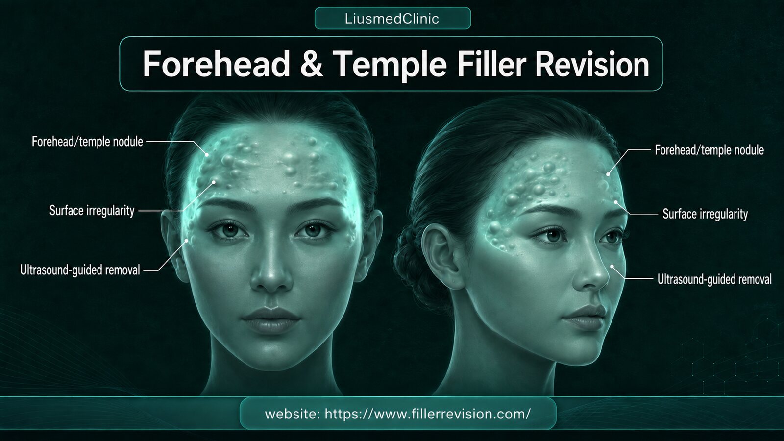
Common Symptoms
Why Forehead Lumps Are Especially Hard to Treat
The forehead skin is thin and sits right against bone, so the slightest unevenness shows immediately; on top of that the region is dense with vessels and nerves, and a lump can lodge at varying depths. These factors together make forehead and temple removal among the most demanding on the whole face — the key is rarely how big the lump is, but knowing exactly which layer it sits in and how close it runs to a vessel. Why lumps form, how the materials differ, migration, and prevention are pre-injection education topics, gathered in the further reading below.
Why Traditional Treatments Fail
Treating Blind Invites Trouble
Pushing, squeezing, or dissecting blindly — without localizing, without seeing clearly — easily means one cut too many into healthy tissue, or one piece left behind that keeps flaring; in the vessel-dense forehead and temple the risk is higher still. Collagen stimulators and permanent materials can't be dissolved anyway and can only be physically removed — but only once the layer, depth, and vessel positions are seen clearly, not hunted for by feel.
“In this region I always look at every spot on ultrasound before I touch anything. Seeing clearly first, then removing, matters more than removing fast.”
Dr. LiuForehead Removal Is Won on Localization and Smoothness
Ultrasound-Guided Pinhole Micro-Extraction
With the forehead and temples, the hard part isn't digging the material out — it's removing it cleanly while keeping the surface smooth. On thin skin over bone nothing can hide, so localization and judgment matter more than anything.
Clean Removal
Collagen stimulators and permanent materials that won't dissolve are physically removed in full under ultrasound guidance — no residue left to keep flaring.
Even Result
After removal on thin-skinned areas the surface is kept as smooth as possible, without creating new dents or waviness.
Precise, Not Excessive
Like fat grafting in reverse — taking only what should come out, protecting the surrounding healthy tissue and this region's dense vessels and nerves.
Precise Removal Under Ultrasound Guidance
The first step in removing a lump isn't the scalpel — it's letting ultrasound see it clearly. We confirm the range, depth, and distribution of every deposit, mark them, plan the path, and only then use a needle-sized entry to take out just what genuinely needs treating, sparing healthy tissue and vessels as much as possible. It's what I always say: not cutting the face open to hunt for lumps, but seeing clearly first, then working through the smallest entry.
Ultrasound localization (draw the lump map)
Marking & path planning
Micro-pinhole removal
Confirm no residue
See It Clearly, Then Remove It
A lump map made for one face
The first step in removing a lump isn't the scalpel — it's letting ultrasound see it clearly first. The lines on this image aren't drawn by hand; each marks a range, depth and distribution confirmed point by point on ultrasound.
Collagen-stimulating materials provoke a strong tissue response and are often wrapped, layer by layer, in a fibrous capsule. The hard part is rarely the size of the lump — it's not knowing where it hides. Dissecting and digging blindly risks one cut too many, one piece left behind, or injury to healthy tissue.
So our order is: scan and localize, then mark, plan the path, and only then use the smallest possible entry to treat what actually needs treating.
Three Months Later, the Same Face
The patient on this map had Ellansé in her forehead. It had been hard lumps for two years, and neither hyaluronidase nor a saline flush had shifted it. After the lumps themselves were removed through pinhole openings, following the map, this is her three months after repair: the same person, the same setting.
Why wait three months to take the photos?
Photograph a forehead right after a flush, or right after a procedure, and it often looks flat, because fluid and swelling in the tissue are temporarily propping the irregularities up. With lumps already encased in collagen, the unevenness usually comes back soon after that fluid is absorbed. What remains once the swelling has settled and the fluid is gone is the real result, which is why the post-treatment photos we use to judge a result are taken at three months.
Not every lump can be removed completely; the goal is to smooth the irregularities and bring back a natural contour. Photos used with the patient's consent. This is an individual case and does not represent results for everyone.
Common Questions
Do forehead filler lumps always need open surgery?
Is it true you'd leave five scars on the face — one each for forehead, temple, tear trough, cheek, and nasolabial fold?
Can collagen-stimulator lumps (Sculptra, Ellansé) in the forehead be removed?
My temple filler has migrated toward the brow tail — can that be treated?
Why is ultrasound essential before removal?
Will forehead removal leave a scar?
Forehead Filler: Understand Before You Decide
Cause, material choice, and prevention are covered on our filler-revision specialty site; removal and repair are performed here.
Not sure what is going on? Write it down and ask.
Describe what was injected, when, and what you are seeing now. Dr. Ta-Ju Liu replies in the thread with an educational explanation of what it might be, what needs to be told apart, and which signs mean you should be seen soon. Posting can be anonymous, and other patients with the same problem can read the answer too.
Replies are general health education, not a diagnosis, and do not replace an in-person examination.