14cm Giant Lipoma Removed Through 1.5cm Incision: Minimally Invasive Case Study
Patient Background
A 55-year-old male patient discovered a lump on his right shoulder over 5 years ago. Recently, the mass had grown significantly, affecting his appearance and daily clothing choices. The patient consulted multiple medical facilities and was told that major surgery with incisions exceeding 10cm would be required.
Patient Concerns
- The shoulder mass had been growing year by year
- The lump was visibly prominent when wearing dress shirts
- Worried about significant scarring after surgery
Pre-operative Assessment
After detailed consultation and physical examination, ultrasound imaging was arranged:
| Examination Item | Result |
|---|---|
| Tumor Location | Right shoulder subcutaneous |
| Tumor Size | 14 x 12 x 5 cm |
| Tumor Depth | Subcutaneous fat layer |
| Boundary | Clear, no invasion of deep tissues |
| Preliminary Diagnosis | Benign lipoma |
Ultrasound imaging showed a homogeneous hypoechoic mass with clear boundaries, consistent with typical lipoma characteristics.
The lateral view clearly shows the degree of protrusion of the mass, with the tumor significantly affecting the shoulder contour.
Surgical Plan
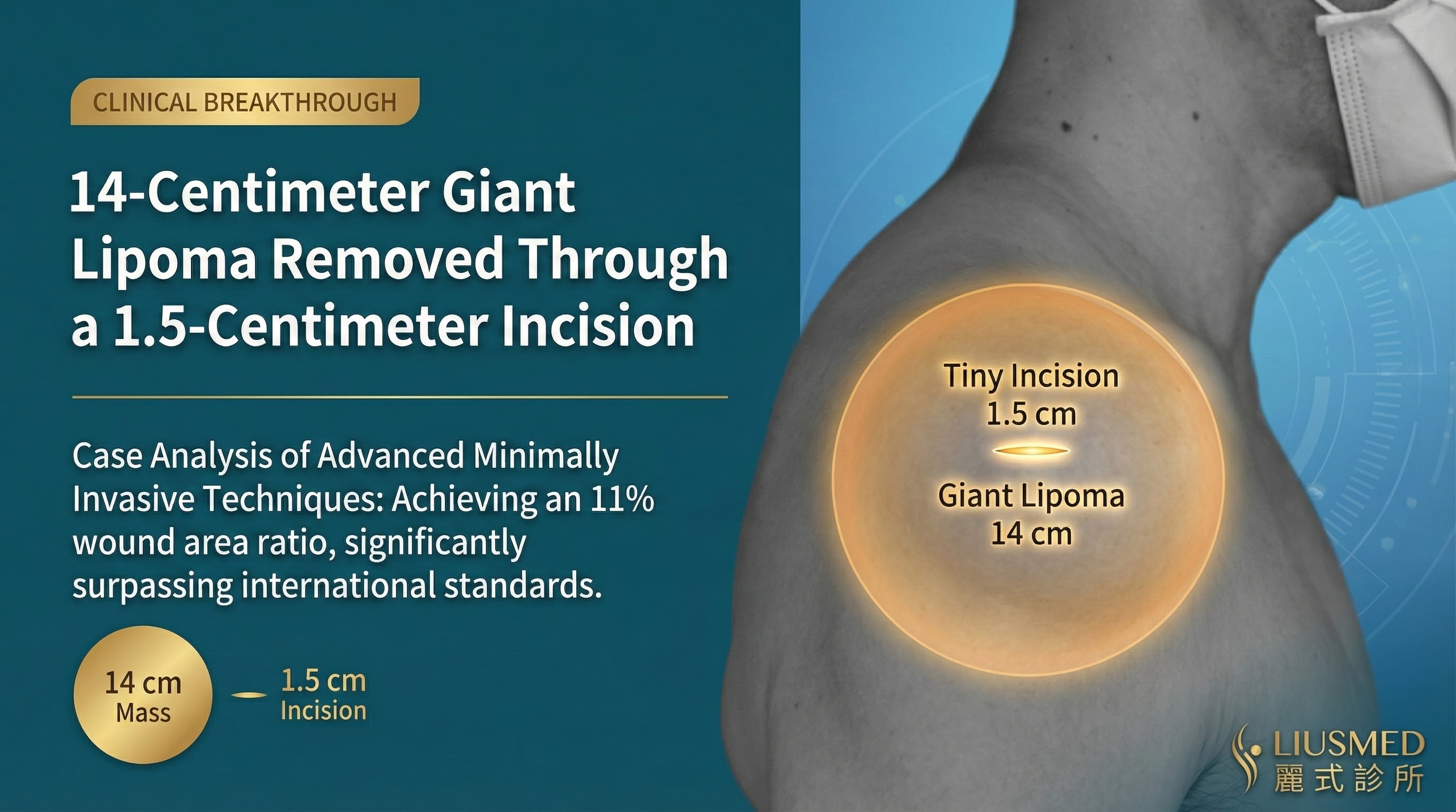
Based on Liusmed Clinic's refined "<20% Minimally Invasive Technique," we assessed that this case could be completed through an approximately 1.5cm incision, with an estimated wound-to-tumor ratio of 11%.
Why Choose Minimally Invasive Surgery?
Traditional surgery for a 14cm lipoma typically requires:
- Equal-length incision: More than 14cm wound
- Extensive dissection: Increased bleeding and tissue damage
- Long recovery time: Requires more rest and healing time
Our minimally invasive technique achieves:
- Ultra-small incision: Only 1.5cm needed
- Precise dissection: Minimized surrounding tissue damage
- Quick recovery: Normal activities resume the next day
Surgical Procedure
The surgery was performed under local anesthesia, taking approximately 45 minutes.
Surgical Steps
- Marking and positioning: Ultrasound-assisted confirmation of tumor location and extent
- Incision design: 1.5cm incision designed along skin creases
- Subcutaneous dissection: Precise dissection using specialized instruments
- Tumor extraction: Complete removal of the 14cm lipoma
- Wound closure: Cosmetic suturing to minimize scarring
Throughout the procedure, the patient remained conscious with no significant pain. The patient was able to walk out of the clinic immediately after surgery.
Incision Ratio Analysis
| Item | Value |
|---|---|
| Tumor Diameter | 14 cm |
| Actual Incision | 1.5 cm |
| Incision Ratio | 11% |
Comparison with International Standards
- MOTIF Technique: Incision ratio approximately 33%
- MIE Technique: Incision ratio approximately 25-50%
- Liusmed Minimally Invasive: Incision ratio < 20%
This case achieved an incision ratio of only 11%, far below all international minimally invasive standards!
Surgical Results
The image above shows the post-operative wound photo. Note the small incision of only about 1.5cm. The tumor was completely removed and the appearance is restored to smooth contour.
Post-operative Recovery
Day 1 Post-op
- Mild wound swelling, no significant pain
- Normal activities resumed, no hospitalization required
- Oral pain medication sufficient
Day 7 Post-op
- Swelling subsided, scab formation began
- Follow-up visit for dressing change, wound healing well
Day 14 Post-op
- Sutures removed, wound completely healed
- Minimal scarring, barely visible
- All daily activities can be resumed
Pathology Report
The excised tissue was sent for pathological examination, confirming a benign lipoma with no malignant cells. The tumor was completely excised with clean margins.
Doctor's Summary
This case fully demonstrates the advantages of our refined minimally invasive technique. For giant lipomas, traditional surgery often requires equal-length incisions, which not only affects aesthetics but also increases recovery time.
Through over 20 years and tens of thousands of surgical cases, we are able to:
- Precise assessment: Detailed preoperative planning ensures surgical safety
- Minimally invasive execution: Minimal incision achieves maximum results
- Complete excision: Ensures no tumor remnants, reducing recurrence rate
- Aesthetic recovery: Concealed, small wounds that are nearly invisible after healing
If you have similar concerns, please schedule a consultation for a personalized treatment plan.
Related Reading

Specialties
Credentials
- Kaohsiung Medical University, School of Medicine
- Attending Physician, Dermatology, Kaohsiung Chang Gung Memorial Hospital
- Attending Physician, Aesthetic Center, Kaohsiung Chang Gung Memorial Hospital
- Visiting Physician, Dermatology, Xiamen Chang Gung Hospital
- Visiting Physician, Aesthetic Center, Xiamen Chang Gung Hospital
"For every surgery, I strive to achieve a good outcome through a small incision and refined technique. Minimally invasive surgery is not just a technique — it's a commitment of respect to every patient."
Want to learn more?
Schedule a consultation for professional evaluation and advice