Minimal-Incision Fat Grafting: Natural Results, Less Swelling
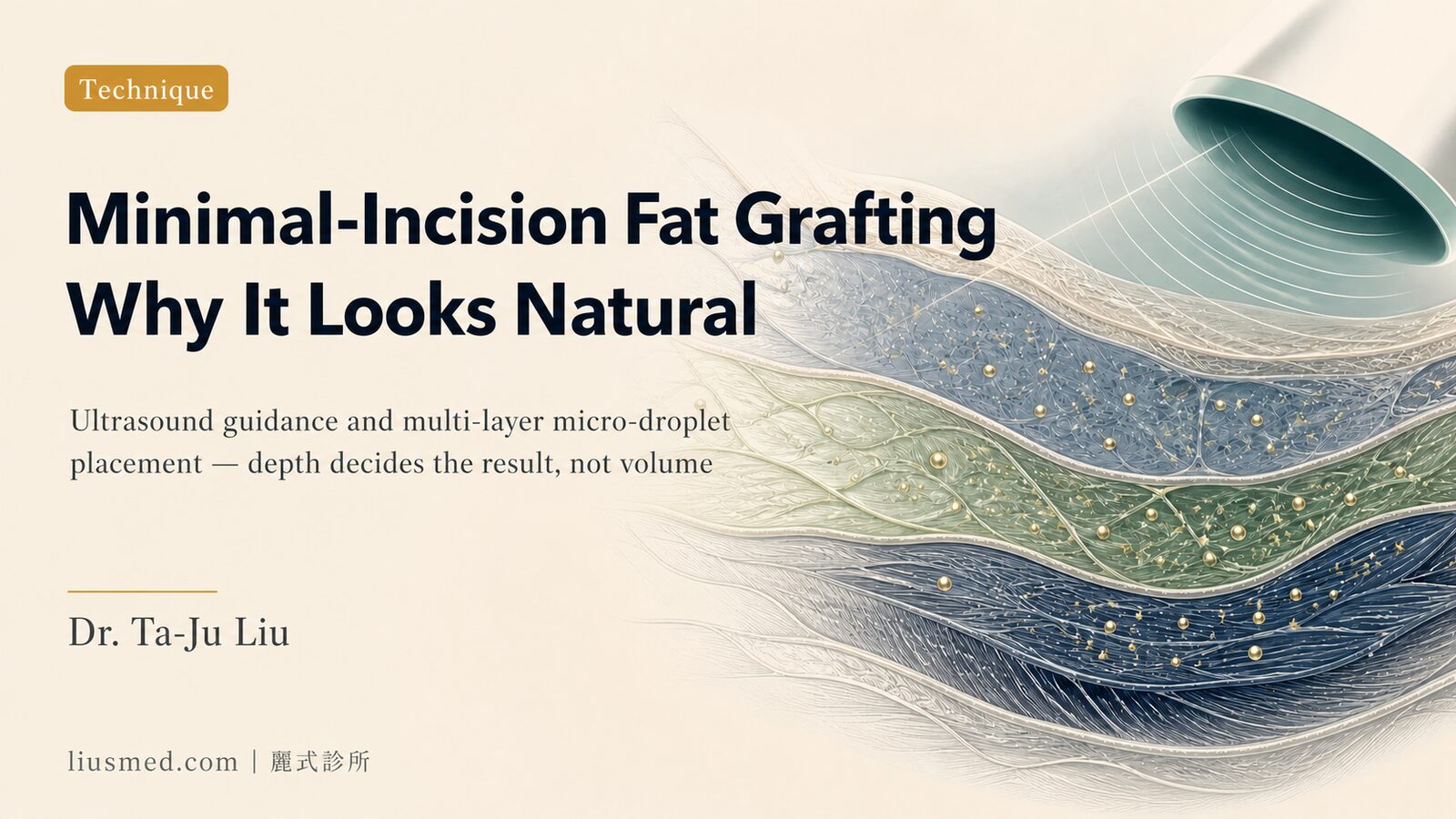
Some patients swell for three months after a fat transfer. Others find that once the swelling resolves, one area looks slightly overfilled, a small mound where the tissue should be smooth. Neither outcome is an inherent flaw of fat grafting. In most cases, both trace back to specific technical choices made during the procedure.
"Minimal incision" in fat grafting refers to more than the length of the access cut. It describes an approach to every stage (harvest, purification, and injection) where reducing tissue trauma is the guiding principle. Each stage has a measurable effect on swelling duration, fat distribution, and long-term survival.
Where Does the Swelling Come From?
Post-operative swelling in fat grafting has two distinct sources: trauma at the harvest site, and the tissue response at the injection site.
During harvest, a larger cannula with higher vacuum pressure damages more capillaries and connective tissue. That means more local bleeding, more fluid accumulation, and a longer recovery. At the injection site, depositing a large volume of fat into a single plane puts more demand on the surrounding tissue than it can immediately support; the inflammatory response is stronger, resorption of excess fluid takes longer, and the blood supply cannot reach the fat cells in the center of a large deposit, increasing the risk of cell death.
Minimal-incision fat grafting addresses both. The goal is not just a smaller cut; it is reducing the total tissue load across the entire procedure.
Harvest Technique: Cannula Size, Vacuum Pressure, and Ultrasound Assistance
Traditional liposuction for fat harvesting often uses cannulas of 3.5–4.5 mm or larger. Efficiency is high, but so is the shear force on fat cells and surrounding structures. Fine-cannula harvest (typically around 2–3 mm) extracts intact fat lobules with less mechanical disruption, and the harvest site recovers with less bruising and swelling.
Ultrasound-assisted liposuction (UAL) adds another layer: sound energy selectively emulsifies and loosens fat cells before removal, allowing extraction through a fine cannula with less physical force. The ultrasound targets fat preferentially, reducing collateral trauma to blood vessels, nerves, and connective tissue. Patients typically notice less bruising at the donor area as a result.
Key point: Fine-cannula, low-pressure harvest reduces trauma at the donor site; less damage to the tissue means less swelling at the harvest location, independent of how much fat is taken.
Purification: Protecting Cell Viability Between Harvest and Injection
Once harvested, fat requires processing before injection: blood, tumescent fluid, oil, and fragmented cells need to be separated. Traditional centrifugation at high G-forces can rupture fat cells; aggressive washing and pressure filtration can also reduce viability if not handled carefully.
The minimal-incision approach prioritizes low-force, low-temperature, and speed: fat cell viability decreases the longer cells remain outside the body, so the time from harvest to injection should be minimized. Gentle separation by gravity sedimentation or low-speed centrifugation preserves the highest proportion of viable cells, which are then the ones that survive and integrate after injection.
Multi-Level Injection: Why Layered Placement Produces Natural Results
This is where the visual outcome is most directly determined.
Depositing a large bolus of fat into a single tissue plane creates a localized concentration of cells that the surrounding blood supply cannot adequately reach. Central fat cells are poorly perfused, die, and can form calcified nodules or firm lumps, one of the most common technical causes of calcification and nodule formation after fat grafting.
Multi-level injection distributes fat across multiple depth planes (the superficial fat compartment, the deep fat pad, and sometimes the fascial layer), depositing only a small amount per pass as the cannula is withdrawn. Each small deposit is surrounded by tissue on all sides, so blood vessels can grow in from multiple directions. Survival rates improve, and the result looks and feels like natural tissue rather than an added layer.
Key point: The volume deposited per pass determines how evenly the fat distributes and how much survives. Small amounts, multiple levels, slow withdrawal: these are the technical variables that produce a natural, non-lumpy result.
Ultrasound-Guided Injection: Knowing Where the Tip Is
Real-time ultrasound during fat injection is rarely discussed but technically significant.
The face has a high density of blood vessels. A minor deviation in needle angle during deep injection can introduce fat into a vessel, causing embolism. For high-risk areas like the tear trough or the deep nasolabial fold, ultrasound imaging allows the surgeon to confirm the needle tip position in real time, verify that fat is being deposited in the target tissue plane, and avoid intravascular placement.
This is the same principle underlying facial fat grafting at our clinic: visualization throughout the procedure, not just at the planning stage.
Traditional vs. Minimal-Incision Fat Grafting: Technical Comparison
| Technical Stage | Traditional Approach | Minimal-Incision Approach |
|---|---|---|
| Harvest cannula size | Larger (≥3.5 mm) | Fine (approx. 2–3 mm) |
| Vacuum pressure | Higher | Low pressure |
| Ultrasound assistance (harvest) | Variable | UAL to loosen fat first |
| Purification method | High-G centrifuge or pressure filtration | Gravity sedimentation or low-speed spin |
| Injection method | Single plane, larger volumes | Multi-level micro-droplet |
| Ultrasound guidance (injection) | Uncommon | Real-time at high-risk areas |
| Post-operative swelling | 1–3 months variable | Peak swelling period typically shorter |
⚠️ Outcomes vary by individual tissue characteristics, volume transferred, and treatment area. The above reflects technical differences in approach; individual results differ.
Survival Rate, Swelling Timeline, and Natural Integration
Fat survival is not a fixed percentage. It depends on cell integrity during harvest, time between harvest and injection, blood supply density at the injection site, and the size of each deposit. Minimal-incision technique has a positive effect on each of these variables; less initial trauma means a lighter inflammatory response, and the body integrates the fat more smoothly.
For a detailed look at the specific factors influencing fat survival, key factors in autologous fat survival rate covers the technical aspects worth understanding before a procedure. The week-by-week swelling timeline and what to expect during recovery are covered in fat grafting swelling and recovery timeline.
Common Questions
Will the access incisions leave visible scars?
Harvest incisions are typically 3–4 mm, under 0.5 cm in length, and rarely leave noticeable scars. Injection on the face follows natural skin lines or concealed entry points with no additional incisions.
Is there enough fat available from a fine cannula?
Facial fat grafting typically requires 5–30 mL depending on area and scope, well within what a fine-cannula harvest can provide. Larger-cannula approaches are only relevant for body contouring volumes.
Three months after surgery and swelling still present: is this a failure?
Not necessarily. Fat integration takes time, and initial firmness or mild swelling is part of the normal process. Localized, persistent hard nodules warrant a follow-up evaluation to rule out calcification or other concerns.
Can energy devices like radiofrequency or HIFU be used after fat grafting?
Allow approximately three months for fat to stabilize before applying strong energy treatments directly over the grafted area. Whether combination procedures are appropriate can be assessed at a follow-up consultation.
To understand which facial areas are well-suited to fat grafting and how different sites compare, facial areas suitable for autologous fat grafting provides a region-by-region breakdown. To assess whether minimal-incision fat grafting is appropriate for your specific situation, schedule a consultation. Facial fat pad distribution and volume loss patterns differ considerably from person to person, and any grafting plan needs to be designed around individual anatomy.

Specialties
Credentials
- Kaohsiung Medical University, School of Medicine
- Attending Physician, Dermatology, Kaohsiung Chang Gung Memorial Hospital
- Attending Physician, Aesthetic Center, Kaohsiung Chang Gung Memorial Hospital
- Visiting Physician, Dermatology, Xiamen Chang Gung Hospital
- Visiting Physician, Aesthetic Center, Xiamen Chang Gung Hospital
"For every surgery, I strive to achieve a good outcome through a small incision and refined technique. Minimally invasive surgery is not just a technique — it's a commitment of respect to every patient."
Recovery after any procedure needs peer support too


Want to learn more?
Schedule a consultation for professional evaluation and advice