
Collagen Stimulator Nodules (Sculptra/Ellansé)
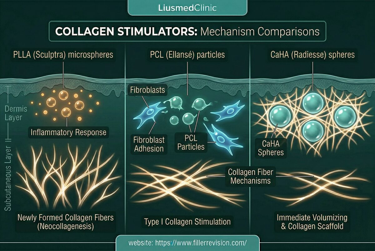
Biostimulatory fillers—including poly-L-lactic acid (Sculptra), polycaprolactone (Ellansé), and calcium hydroxylapatite (Radiesse)—are designed to stimulate collagen production but cannot be dissolved by any enzyme. When complications occur, they manifest as hard, palpable nodules or granulomatous reactions that can appear months to years after injection. Published nodule incidence rates range from 1-10% depending on the product and technique. These complications are particularly challenging because the particles become embedded within the body's own newly-formed collagen matrix, creating a composite material that resists all non-surgical interventions.
Three Core Strengths of Liusmed Revision

Clean Removal
Stubborn masses are removed completely so residual tissue does not keep flaring.

Even Result
After removal the surface stays smooth, without new dents or waviness.

Precise, Not Excessive
Like fat grafting in reverse — the amount and location removed are judged precisely.
Common Symptoms
Particle Aggregation & Immune Response
Biostimulator complications arise from two primary mechanisms. First, improper dilution, inadequate mixing, or injection into the wrong tissue plane causes particle aggregation—clusters of microspheres that trigger an exaggerated foreign body response. The body attempts to wall off these clusters with dense fibrous tissue, forming palpable nodules. Second, in susceptible individuals, the immune system mounts a granulomatous reaction—recruiting macrophages, giant cells, and inflammatory mediators that form an organized inflammatory mass around the particles. Unlike HA complications, these cannot be chemically dissolved; the particles persist within a collagen-fibrous matrix that only responds to physical intervention.
Why Traditional Treatments Fail
Why Conservative Treatments Fail
Since biostimulators are not hyaluronic acid, enzymatic dissolution has zero effect. Intralesional steroid injections can temporarily reduce inflammation and nodule size, but they do not remove the foreign body trigger—leading to recurrence once treatment is stopped. Prolonged steroid use carries significant risks: fat atrophy causing visible skin depressions, skin thinning, telangiectasia, and hypopigmentation. Other injectable agents can soften early-stage inflammatory nodules but have limited efficacy against mature, encapsulated nodules where dense scar tissue has already formed around the particle clusters. The fundamental problem remains: without physically removing the foreign particles, the body's immune response will continue indefinitely.
“With biostimulator nodules, the key is preserving the surrounding fat while removing only the foreign particles. Steroids melt everything—including the healthy tissue you want to keep.”
Dr. LiuWhen Nothing Can Dissolve It, Physics Is the Only Answer
Ultrasound-Guided Pinhole Micro-Extraction
Biostimulator particles — PLLA, PCL, CaHA — are immune to every enzyme and medication available. The only path to resolution is physical extraction. But the critical challenge isn't just removing the foreign particles; it's removing them while preserving the patient's own fat and collagen that surround them.
No Chemical Shortcut Exists
Unlike HA, these materials have no dissolving agent. Steroids suppress the inflammation but leave the trigger intact, guaranteeing that the nodule will return once the steroid effect wears off.
Protect the Host, Extract the Foreign
The surrounding fat pad and collagen matrix are the patient's own healthy tissue. Aggressive approaches sacrifice this tissue unnecessarily; precision extraction preserves it.
Steroid-Free Resolution
Avoiding repeated steroid cycles prevents the iatrogenic complications — fat atrophy, skin thinning, visible depressions — that often become worse than the original nodule.
Pinhole Micro-Extraction
Our approach targets the root cause: physical removal of the foreign particle clusters. Using ultrasound to precisely locate each nodule, we perform micro-dissection to create a safe separation plane that pushes neurovascular structures aside, isolating the nodule from surrounding healthy tissue. We then mechanically fragment the compacted material into extractable pieces. The fragments are aspirated through a pinhole entry point. This achieves definitive resolution without the tissue damage, scarring, or fat atrophy associated with open surgery or repeated steroid injections.
Ultrasound Localization
Micro-dissection
Mechanical Fragmentation
Aspiration & Verification
See It Clearly, Then Remove It
A lump map made for one face
The first step in removing a lump isn't the scalpel — it's letting ultrasound see it clearly first. The lines on this image aren't drawn by hand; each marks a range, depth and distribution confirmed point by point on ultrasound.
Collagen-stimulating materials provoke a strong tissue response and are often wrapped, layer by layer, in a fibrous capsule. The hard part is rarely the size of the lump — it's not knowing where it hides. Dissecting and digging blindly risks one cut too many, one piece left behind, or injury to healthy tissue.
So our order is: scan and localize, then mark, plan the path, and only then use the smallest possible entry to treat what actually needs treating.
Three Months Later, the Same Face
The patient on this map had Ellansé in her forehead. It had been hard lumps for two years, and neither hyaluronidase nor a saline flush had shifted it. After the lumps themselves were removed through pinhole openings, following the map, this is her three months after repair: the same person, the same setting.
Why wait three months to take the photos?
Photograph a forehead right after a flush, or right after a procedure, and it often looks flat, because fluid and swelling in the tissue are temporarily propping the irregularities up. With lumps already encased in collagen, the unevenness usually comes back soon after that fluid is absorbed. What remains once the swelling has settled and the fluid is gone is the real result, which is why the post-treatment photos we use to judge a result are taken at three months.
Not every lump can be removed completely; the goal is to smooth the irregularities and bring back a natural contour. Photos used with the patient's consent. This is an individual case and does not represent results for everyone.
Common Questions
Are collagen-stimulator lumps in the forehead or temple handled differently?
Can Sculptra lumps go away on their own?
Is Ellanse harder to remove than Sculptra?
Need surgery?
Can it come back?
Will there be a dent where the nodule was?
How long after injection do lumps appear?
Why didn't steroid injections dissolve my Sculptra lump?
Area Focus: Forehead & Temple
Cause, material choice, and prevention are covered on our filler-revision specialty site; removal and repair are performed here.
Composition, brands, and removability — compiled on our filler-revision specialty site.
Not sure what is going on? Write it down and ask.
Describe what was injected, when, and what you are seeing now. Dr. Ta-Ju Liu replies in the thread with an educational explanation of what it might be, what needs to be told apart, and which signs mean you should be seen soon. Posting can be anonymous, and other patients with the same problem can read the answer too.
Replies are general health education, not a diagnosis, and do not replace an in-person examination.