Case Analysis: Nose Filler Displacement — Extraction and Nasal Contour Reconstruction Record
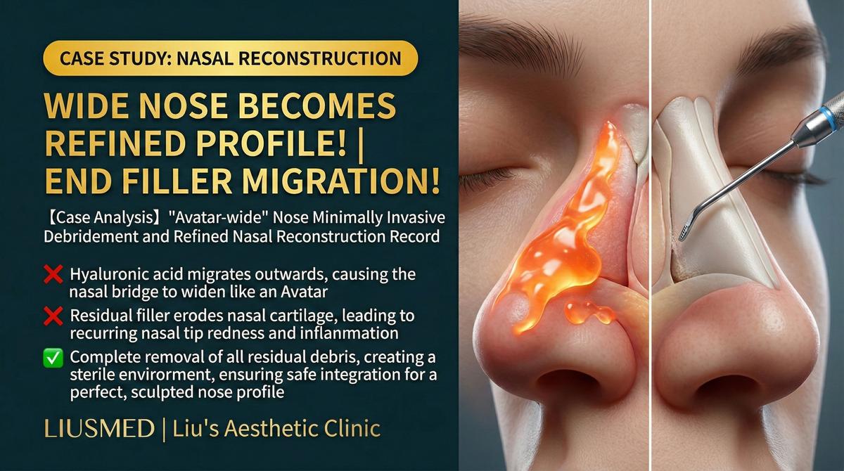
Case Scenario
Patient Background: A woman in her 30s who received hyaluronic acid injection rhinoplasty at a cosmetic clinic approximately 18 months prior. The initial result was satisfactory, with a straighter nasal bridge. However, approximately 4 months post-injection, the patient noticed asymmetry along the nasal dorsum — one side appeared wider than the other, and the filler could be felt in positions different from the original injection sites.
Treatment History:
- First noticed left-sided nasal dorsum prominence approximately 4 months post-injection
- Returned to the original clinic and was advised that "mild asymmetry will resolve naturally"
- Waited an additional 3 months, but asymmetry worsened rather than improved
- Underwent hyaluronidase injection at the original clinic, but results were uneven — some areas over-dissolved while others remained inadequately treated
- Post-dissolution nasal contour became more irregular with surface bumps and depressions
- Found information about ultrasound-guided precision extraction through online research
Presentation at Consultation:
- Visible nasal dorsum asymmetry with left-sided prominence
- Palpation revealed irregular distribution of residual filler in multiple locations
- Several areas of surface irregularity on the nasal dorsum (evidence of uneven hyaluronidase treatment)
- Slight tip rounding with loss of original refinement
- Patient's dissatisfaction with nasal appearance had significantly impacted social confidence
Deep Analysis
Root Cause Analysis
| Aspect | Finding |
|---|---|
| Displacement cause | Thin nasal skin with limited tissue space; filler affected by facial expression muscle movement and gravity, resulting in lateral spread |
| Initial injection assessment | Injection plane likely too superficial, making filler more susceptible to tissue movement |
| Hyaluronidase issues | Non-ultrasound-guided blind hyaluronidase injection caused uncontrolled dissolution — simultaneous over-dissolution in some areas and persistent residue in others |
| Ultrasound findings | Multiple irregular deposits of residual HA across the nasal dorsum, some displaced over the alar cartilage |
| Tissue condition | Mild fibrotic reaction from repeated injection and dissolution cycles |
Key Insight: The nose is a high-risk zone for filler displacement. Nasal skin is thin, tissue space is limited, and the area is subject to continuous movement from facial expressions. Once displacement occurs, non-guided hyaluronidase injection often fails to address the problem precisely — it may over-dissolve in areas that don't need treatment while inadequately reaching the actual displaced material. Ultrasound guidance is the critical tool for managing these complex nasal filler complications.
Related reading: How to Handle Nose Filler Displacement
Doctor's Perspective
"Nasal filler displacement is a remarkably common complication we encounter in our clinic. This patient's situation represents a classic treatment dilemma: after filler displacement occurs, non-guided hyaluronidase injection actually makes the problem more complex. Ultrasound examination clearly showed the distribution of residual HA (Hyaluronic Acid — sugar molecule naturally in skin, holds water) — it was no longer concentrated along the nasal midline but had spread laterally to the left, with some material having migrated to unexpected positions.
The key principles for managing these cases are: first, use ultrasound for a complete assessment of the precise location and extent of residual filler; second, develop a zone-by-zone treatment strategy — perform targeted extraction of clearly identified deposits rather than blind dissolution; third, protect the nose's normal tissue architecture during extraction, particularly the delicate anatomical layers above the dorsal cartilage perichondrium."
Treatment Plan and Process
Preoperative Planning
| Planning Element | Details |
|---|---|
| Ultrasound assessment | Complete scanning of nasal dorsum, alae, and tip; marking all residual filler locations and depths |
| Vascular mapping | Confirming dorsal nasal artery course to ensure safe extraction pathways |
| Zone planning | Dividing the nose into four treatment zones: radix, upper dorsum, lower dorsum, and tip |
| Entry point selection | Selecting concealed positions inside the nostril and along the lateral nasal dorsum for micro-incisions |
Surgical Process
- Precise anesthesia: Ultrasound-guided nasal nerve block and local infiltration, plus gentle pain-relief anesthesia
- Zone-by-zone scan confirmation: Pre-operative re-confirmation of filler locations in each zone
- Priority treatment of most displaced area: Beginning with the left dorsal displacement accumulation zone
- Real-time ultrasound guidance: Operating entirely under ultrasound visualization to ensure instrument accuracy
- Stepwise precision extraction: Using micro-instruments to extract residual filler incrementally, avoiding excessive tissue disruption
- Immediate assessment: Scanning after each extraction to confirm results
- Symmetry verification: Post-extraction comparison of bilateral nasal dorsum symmetry
Extraction Results
Multiple irregularly distributed residual HA deposits were successfully extracted, including partially degraded old filler masses. Extracted material ranged from translucent gel-like consistency to opaque, denatured masses — reflecting the different states resulting from varying injection periods and prior hyaluronidase treatment. Post-operative ultrasound confirmed significant reduction in residual material across the nasal dorsum.
Key Patient Notes
Special Considerations for Nasal Extraction
| Characteristic | Explanation |
|---|---|
| Delicate nasal anatomy | The nose's anatomical layers are thin and precise, requiring exceptional care during manipulation |
| Prominent swelling | The nose has rich blood supply; post-operative swelling may be more noticeable than other areas |
| Appearance requires recovery time | The nose may look suboptimal initially due to swelling; patience is essential |
| Possible follow-up adjustment | If minor depressions remain after extraction, assessment for small-volume precision filling may be needed |
| Long-term follow-up matters | Regular post-operative follow-up appointments are recommended to monitor recovery and stability |
Recovery Expectations
| Timeline | Expected Condition |
|---|---|
| Days 1-3 | Peak swelling; mild bruising around the nose possible |
| Week 1 | Swelling begins to subside; bruising gradually fades |
| Weeks 2-4 | Nasal contour progressively clarifies; symmetry improvement becomes apparent |
| Month 2 | Tissue continues to soften and remodel; approaching final result |
| About 3 months | Close to the final result (varies by person); assessment for any needed fine-tuning |
Key Insight: Recovery after nasal filler extraction requires particular patience. The nose is the visual focal point of the face — even slight swelling is easily noticeable. It is important to understand that the immediate post-operative appearance is not the final result. As swelling resolves and tissue remodels, the nasal shape will progressively return to a more natural state.
Clinical Takeaways
- Nasal filler displacement warrants early evaluation — waiting for "natural absorption" typically only delays treatment; displaced filler does not return to its original position on its own
- Non-guided hyaluronidase injection carries high risk in the nose — the nose's delicate structure makes blind dissolution likely to produce uneven results
- Ultrasound is foundational for nasal filler problem management — precise localization enables precise treatment
- Post-extraction nasal assessment needs time — additional procedures should not be rushed during the swelling phase
- Choose a physician with nasal anatomy expertise — the nose's fine structure demands deep anatomical knowledge
If you are experiencing nasal filler complications, schedule a consultation for a precision ultrasound assessment.
Related reading:
- How to Handle Nose Filler Displacement
- Filler Lump Extraction Technique
- Filler Repair Evaluation Process
Related Services

Specialties
Credentials
- Kaohsiung Medical University, School of Medicine
- Attending Physician, Dermatology, Kaohsiung Chang Gung Memorial Hospital
- Attending Physician, Aesthetic Center, Kaohsiung Chang Gung Memorial Hospital
- Visiting Physician, Dermatology, Xiamen Chang Gung Hospital
- Visiting Physician, Aesthetic Center, Xiamen Chang Gung Hospital
"For every surgery, I strive to achieve a good outcome through a small incision and refined technique. Minimally invasive surgery is not just a technique — it's a commitment of respect to every patient."
Recovery after any procedure needs peer support too


Want to learn more?
Schedule a consultation for professional evaluation and advice