Full-Face Ultrasound Filler Audit: Seeing the Truth Beneath Your Skin
Why Do You Need a Full-Face Ultrasound Filler Audit?
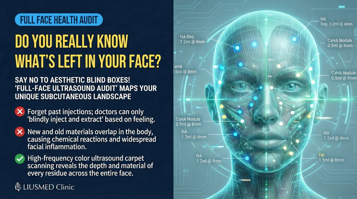
In an era of increasingly widespread filler injections, many people face a common problem: they are uncertain what exactly is in their face. After years of injections by multiple physicians, various types and layers of filler may have accumulated beneath the skin — and the patient often can no longer recall the details.
The full-face ultrasound filler audit is a systematic assessment service designed to solve exactly this problem.
Key Insight: A filler audit is not about "checking if there is a problem" — it is about "seeing the complete truth beneath the skin." Many patients discover after their audit that they have far more filler than they imagined, and in different locations than they remembered.
Who Needs a Full-Face Filler Audit?
| Candidate | Situation |
|---|---|
| Multiple injection history | Years of injections by various physicians; uncertain what remains |
| Planning revision | Wants to remove or revise filler; needs complete pre-operative assessment |
| Experiencing symptoms | Feels facial abnormalities (lumps, asymmetry, pain, etc.) |
| Considering re-injection | Wants to add filler to an existing base; needs to confirm safety |
| Uncertain injection history | Unsure of past materials or injection locations |
| Routine monitoring | Has known filler and wants periodic status confirmation |
What Does a Full-Face Ultrasound Audit Include?
Scanning Regions
A complete full-face filler audit covers all of the following zones:
| Region | Scanning Focus | Common Findings |
|---|---|---|
| Forehead | Frontal to glabellar area | Filler migration to glabella, surface irregularity |
| Temples | Superficial temporal artery course | Deep filler deposits, vascular compression |
| Glabella | Wrinkle injection zone | Unknown material residuals |
| Periorbital | Tear trough, eye bag area | Tyndall effect causes, deep residuals |
| Nose | Dorsum, tip, alar | Filler migration, vascular compression risk |
| Cheeks / Malar | Mid-face region | Overfilling, asymmetry |
| Nasolabial folds | Nasolabial groove | Multiple injection accumulation |
| Lips | Upper/lower lip, perioral | Lumps, unnatural lip shape |
| Chin | Mentum | Filler migration |
| Jawline | Mandibular margin | Contour indistinctness |
Assessment Items
The ultrasound evaluation for each region includes:
| Assessment Item | Method | Clinical Significance |
|---|---|---|
| Filler presence | B-mode ultrasound | Confirm whether filler exists |
| Filler type | Echo characteristic analysis | Determine likely material type |
| Filler depth | Depth measurement | Confirm anatomical layer of filler |
| Filler extent | Multi-plane scanning | Confirm three-dimensional distribution |
| Filler condition | Texture and morphology analysis | Assess for aggregation, fibrosis, etc. |
| Vascular relationship | Color Doppler | Confirm filler-to-vessel spatial relationship |
| Complication signs | Comprehensive imaging analysis | Early detection of potential problems |
How Ultrasound Identifies Different Filler Types
Ultrasound Characteristics by Filler Type
| Filler Type | Ultrasound Appearance | Identification Key |
|---|---|---|
| HA (Hyaluronic acid) | Anechoic or hypoechoic | Relatively clear margins, compressible |
| Collagen stimulators (e.g., Sculptra) | Hyperechoic punctate distribution | Scattered hyperechoic granules |
| Silicone / Silicone oil | Hyperechoic with posterior snowstorm artifact | Characteristic "blizzard" pattern |
| PMMA | Hyperechoic with posterior acoustic shadow | Well-defined hyperechoic mass |
| CaHA (Radiesse) | Hyperechoic punctate with acoustic shadow | Calcification-like hyperechoic pattern |
| Autologous fat | Similar echogenicity to surrounding fat | May have fibrous capsule or cysts |
For more on ultrasound filler identification, see Ultrasound Imaging for Filler Identification.
Key Insight: Different filler types produce distinctly different ultrasound images. An experienced physician can determine filler type from these characteristics, even when the patient cannot remember what was injected.
What Does the Audit Report Include?
After completing the full-face ultrasound scan, you receive a detailed audit report:
Report Contents
| Report Item | Description |
|---|---|
| Full-face filler distribution map | Marks location and extent of filler in each region |
| Regional detailed assessment | Ultrasound findings for each scanned zone |
| Filler type inference | Estimated filler types based on ultrasound characteristics |
| Vascular safety assessment | Spatial relationship between filler and critical vessels |
| Risk assessment | Identification of potential issues with risk grading |
| Recommended plan | Suggested next steps (observation / revision / follow-up) |
Possible Action Plans After Audit
| Audit Finding | Recommended Action | Explanation |
|---|---|---|
| Everything normal | Routine follow-up | Recheck every 1–2 years |
| Unknown material found | Further evaluation | Confirm material type and risk |
| Migration detected | Assess revision necessity | Decision based on severity |
| Vascular compression found | Priority treatment | Vascular safety is top priority |
| Fibrosis/Lumps found | Evaluate extraction | Decision based on symptoms and risk |
| Excessive filler | Develop volume reduction plan | Staged extraction |
Clinical Value of Full-Face Ultrasound Audit
Value for Patients
- Right to know: Understand the true state beneath your facial skin
- Safety assurance: Early detection of potential risks
- Decision support: Objective information for treatment planning
- Record establishment: Build a complete filler history record
Value for Subsequent Treatment
- Precise surgical planning: Know what to extract, where, and how deep
- Risk prediction: Pre-identify potential intraoperative risks
- Efficiency improvement: Reduce exploratory time during surgery
- Outcome optimization: More precise extraction leads to better results
Key Insight: A full-face filler audit is the "navigation map" for revision surgery. Just as imaging studies are required before any surgical procedure, a pre-revision ultrasound scan dramatically improves surgical safety and precision.
The Audit Process at Liusmed Clinic
| Step | Content | Duration |
|---|---|---|
| Medical history | Past injection history and current symptoms | 10–15 minutes |
| Clinical examination | Visual and palpation assessment | 5–10 minutes |
| Ultrasound scanning | Systematic full-face ultrasound scan | 30–45 minutes |
| Image analysis | Analysis of ultrasound findings | 10–15 minutes |
| Report explanation | Explaining audit results and recommendations to the patient | 15–20 minutes |
For more on the evaluation process, see Filler Repair Evaluation Process.
Conclusion: Seeing Is the First Step to Making the Right Decision
A full-face ultrasound filler audit is a simple yet enormously valuable assessment service. Whether you are planning revision, considering additional injections, or simply want to understand what lies beneath your facial skin, this examination provides the answers you need.
"See the truth beneath your skin" — this is the first step toward any sound medical decision.
Contact Liusmed Clinic to schedule a full-face ultrasound filler audit.
Related reading: Filler Repair Evaluation Process, Ultrasound Imaging for Filler Identification, Ultrasound Diagnosis and Treatment Golden Standard
Related Services

Specialties
Credentials
- Kaohsiung Medical University, School of Medicine
- Attending Physician, Dermatology, Kaohsiung Chang Gung Memorial Hospital
- Attending Physician, Aesthetic Center, Kaohsiung Chang Gung Memorial Hospital
- Visiting Physician, Dermatology, Xiamen Chang Gung Hospital
- Visiting Physician, Aesthetic Center, Xiamen Chang Gung Hospital
"For every surgery, I strive to achieve a good outcome through a small incision and refined technique. Minimally invasive surgery is not just a technique — it's a commitment of respect to every patient."
Recovery after any procedure needs peer support too
Want to learn more?
Schedule a consultation for professional evaluation and advice