Can You Redo Rhinoplasty Before Residual Nasal Filler Is Fully Cleared?
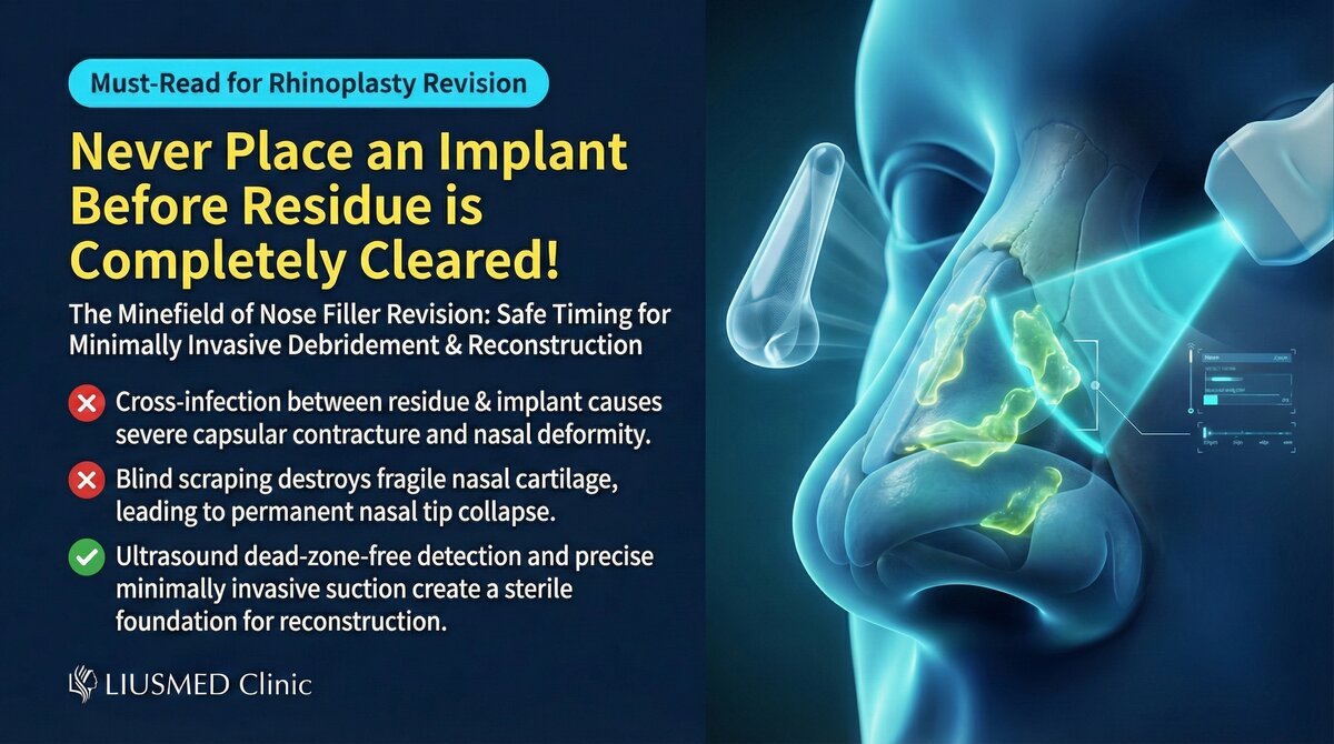
How Residual Filler Impacts Rhinoplasty Reconstruction
Many patients considering rhinoplasty revision overlook a critical prerequisite: whether previous nasal filler has been completely cleared. Residual filler not only interferes with the surgical procedure but may jeopardize the long-term outcome.
Risks of Residual Filler
| Risk Type | Specific Impact | Consequence |
|---|---|---|
| Space occupation | Residual material occupies space meant for implant or cartilage | Incorrect implant position, poor aesthetics |
| Infection risk | Old filler may harbor bacteria or biofilm | Elevated post-operative infection rate |
| Tissue reaction | Residual continues to stimulate foreign body response | Chronic inflammation, capsular contracture |
| Blood circulation | Residual compresses or obstructs local blood flow | Poor tissue healing, implant exposure |
| Unpredictable appearance | Residual overlaps with new implant | Unnatural or asymmetric appearance |
Key Insight: Performing rhinoplasty reconstruction with residual filler in place is equivalent to building a house on an unclean foundation. No matter how skilled the surgical technique, an unstable foundation compromises the structure's durability.
Common Nasal Filler Residual Scenarios
Problems Frequently Found on Ultrasound
| Scenario | Ultrasound Finding | Patient's Usual Assumption |
|---|---|---|
| Post-dissolution residual | Incompletely dissolved HA remaining | Believed dissolution was complete |
| Deep-layer residual | Filler on supraperiosteal plane untreated | Unaware of deep-layer residual |
| Migration and diffusion | Filler spread from bridge to ala or glabella | Only noticed the bridge problem |
| Encapsulation | Filler encased in fibrous tissue | Assumed absence because it was not palpable |
| Mixed materials | Multiple materials from different sessions coexisting | Cannot recall what was previously injected |
For more on nasal filler migration, see Nose Filler Migration.
The Correct Treatment Sequence
Step 1: Comprehensive Ultrasound Assessment
Before deciding on any surgery:
- Ultrasound scan of the entire nasal area: Root, bridge, tip, ala, dorsum
- Identify filler type: Different materials show different ultrasound characteristics
- Measure residual extent: Confirm position, size, and depth of residual material
- Assess tissue condition: Skin thickness, fibrosis degree, vascular distribution
- Create distribution map: Provide navigation basis for extraction surgery
Step 2: Minimally Invasive Filler Extraction
Using ultrasound-guided minimally invasive technique:
| Nasal Area | Extraction Key Points | Special Precautions |
|---|---|---|
| Nasal root | Watch for angular artery | Near the eye, extreme caution required |
| Nasal bridge | Mostly superficial, relatively direct | Watch for skin damage at thin areas |
| Nasal tip | Tight space, delicate structures | Protect tip cartilage support |
| Nasal ala | May contain diffuse filler | Preserve alar cartilage integrity |
Step 3: Wait for Tissue Recovery
Rhinoplasty should not be performed immediately after extraction:
- Minimum wait: 3 months after extraction
- Recommended wait: 6 months after extraction
- Ideal: Tissue fully stabilized, ultrasound confirms no residual
Step 4: Rhinoplasty Reconstruction Assessment
Once tissue has stabilized, evaluate rhinoplasty reconstruction:
- Assess nasal tissue conditions (skin, cartilage, bone)
- Select appropriate reconstruction material (autologous cartilage, implant, etc.)
- Develop a personalized surgical plan
Key Insight: Patience pays off. Performing rhinoplasty after tissue has fully recovered and residual filler clearance is confirmed significantly improves success rates and satisfaction.
Why Not Extract and Reconstruct Simultaneously?
Some patients want to "solve everything at once." However:
| Risk of Simultaneous Approach | Explanation |
|---|---|
| Increased infection risk | Extraction and implantation together complicates infection control |
| Uncertain tissue condition | Post-extraction tissue needs time to stabilize |
| Imprecise planning | Cannot predict exact appearance changes after extraction |
| Uncertain reconstruction space | Residual tissue reaction may alter internal space |
| Doubled revision difficulty | Problems from both procedures compound each other |
Conclusion: Clear First, Then Rebuild
The correct logic for nasal filler revision is first ensure complete clearance of residual material, then proceed with rhinoplasty reconstruction. This sequence may seem to prolong treatment, but it dramatically improves the quality and safety of the final result.
If you are considering rhinoplasty but uncertain whether previous filler has been fully cleared, contact Liusmed Clinic for an ultrasound evaluation.
Related reading: Filler Lump Extraction Technique, Filler Repair Evaluation Process
Related Services

Specialties
Credentials
- Kaohsiung Medical University, School of Medicine
- Attending Physician, Dermatology, Kaohsiung Chang Gung Memorial Hospital
- Attending Physician, Aesthetic Center, Kaohsiung Chang Gung Memorial Hospital
- Visiting Physician, Dermatology, Xiamen Chang Gung Hospital
- Visiting Physician, Aesthetic Center, Xiamen Chang Gung Hospital
"For every surgery, I strive to achieve a good outcome through a small incision and refined technique. Minimally invasive surgery is not just a technique — it's a commitment of respect to every patient."
Recovery after any procedure needs peer support too


Want to learn more?
Schedule a consultation for professional evaluation and advice