Fat Grafted Under the Eye Turned Into a Lump, Then a Stimulator Was Added to Hide It: A Full Record of Removing Five-Year Stacked Nodules
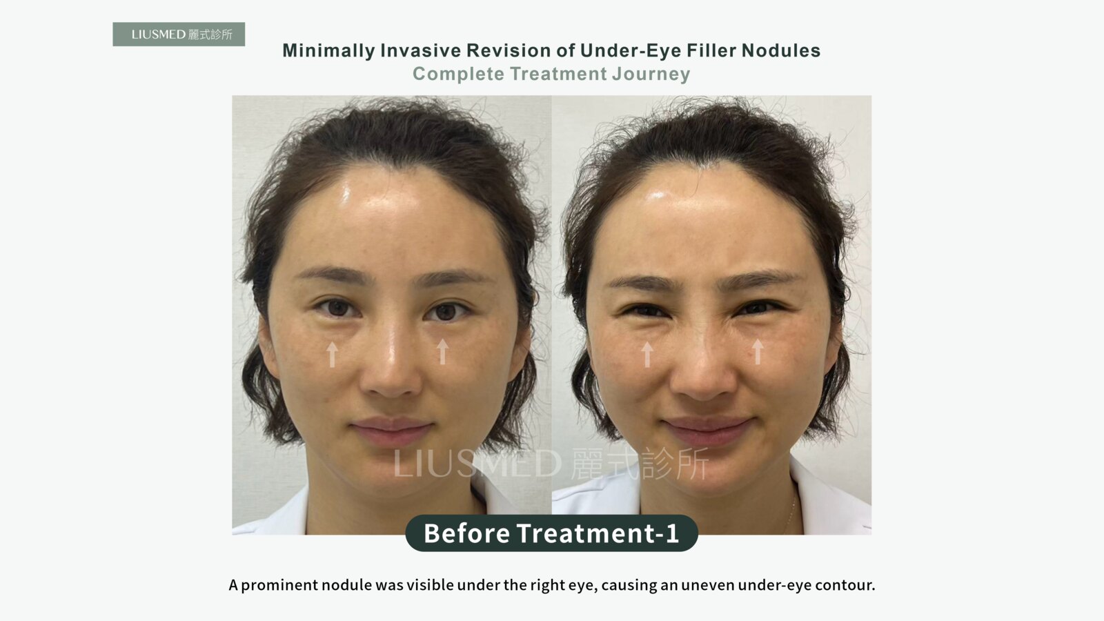
Most case write-ups show you two photographs: before and after. What happened in between, how the surgeon decided where to go in, and what actually came out that day are usually left out.
This one is different. All four stages of this patient's treatment were documented: before, ultrasound marking, the moment of removal, and three months later. I want to use this single case to walk through what handling an under-eye nodule really involves.
How one problem became two
The original request was simple enough: fill the tear troughs.
Fat was grafted under her eyes, and the hollow did fill in. Some weeks later, though, she could feel something firm there, and the bulge was visible from the outside.
What happened next is a path a lot of people go down. If the raised part will not go away, then lift the dip beside it, and the whole thing should look level. So a collagen stimulator was placed next to it.
That injection left a second nodule.
From that point on, the two of them sat under her eye for more than five years.
They showed at rest. They showed far more when she smiled, because the orbicularis muscle contracts and pushes the whole thing forward. Friends kept asking her outright: what happened under your eye, what did you have injected, why does it look like that?
Being asked was not the hard part. The hard part was that she had no answer either. She had wanted it gone for years, and what she kept hearing was either wait and see whether it absorbs, or this needs surgery.
Why adding a little more to cover it rarely works
The most instructive part of this case is that second step, the one that feels intuitive and almost never works.
The under-eye area is already raised, so you add material beside it to bring the surroundings up to the same height and erase the difference. In practice, three things happen.
First, you now have one more thing to deal with. One nodule became two, and the two are made of different materials.
Second, the eye area is not a flat surface. The skin is thin, the orbicularis muscle underneath moves constantly, and material placed here gets pushed into positions you did not plan for. That is part of why removal around the eye is among the most precise work on the face.
Third, thin skin plus raking light means any difference in height gets amplified by its own shadow. You think you have levelled it, and the mirror still says otherwise.
Put plainly: adding material is not how you fix an under-eye bulge.
Stage two: see it first, then decide what to take out
Before doing anything, I scanned the entire under-eye area with high-frequency ultrasound.
That step matters especially here, because there was more than one kind of material to face. A mass formed by grafted fat and a nodule formed by a collagen stimulator usually sit at different depths and in different places. The first tends to be deeper, with borders that blur into the patient's own tissue. The second may sit shallower and stay more contained.
Only once you can see which layer each one is in, and how far it extends, can you mark the position on the skin and decide where the pinhole goes and which direction it travels.
Those lines in the photo are not decoration. They are what the ultrasound found, transferred back onto the surface. Without that step, the rest is done by feel, and around the eye the margin for error next to vessels and nerves is small.
This is the principle we keep coming back to: you have to see it to handle it safely. The same approach is set out in more detail in minimally invasive repair after failed facial fat grafting.
Stage three: the day of removal
The removal was done through a pinhole-sized opening.
The two syringes in that photo hold what actually came out that day. Not an illustration. The material that had been sitting in her face for five years.
We take this photograph because people keep asking the same question: how do I know it really came out? That is the answer.
Stage four: three months later
At the three-month follow-up, the under-eye contour is even again. Same angles, same lighting, before and three months after placed side by side:
The 45-degree view is worth a second look. Straight on, part of the nodule hides in its own shadow. Turn to 45 degrees and oblique light brings out the whole step in the contour, and that edge is hard to miss. At three months, same angle and same light, the edge is no longer there.
To be clear, this is how this particular patient recovered. Appearance after removal, how quickly swelling settles, and how well the skin springs back all vary from person to person, and depend on how much was placed, how long it stayed, and the condition of the skin.
Fat nodules and stimulator nodules are not the same problem
These two get lumped together in conversation, but the logic of handling them differs.
Grafted fat is your own tissue. The portion that survives stays long term; the portion that does not may fibrose or calcify into something you can feel. What makes it awkward is that there is no clean boundary between it and the surrounding native fat, so on ultrasound it has to be distinguished by echo characteristics and border morphology.
A collagen stimulator is foreign material that works by prompting the tissue to lay down collagen. The nodules it forms usually have clearer borders, but because they are the body's response to a foreign body, the tissue around them tightens over time.
So that preoperative scan is not only about location. It is also about judging the state of each material and deciding how each one should be approached. For more on how filler nodules are handled, see filler complication repair; if your situation stems mainly from fat grafting, fat graft complication repair covers that side.
This case is also documented on our filler revision site, from the angle of telling the materials apart and deciding whether a second injection makes sense: fat graft, then a collagen stimulator.
The same approach, mapping with ultrasound first and then removing through pinholes, is documented for the forehead too, at three months: Two years of Ellansé forehead lumps: from ultrasound map to three months after repair.
A bulging upper eyelid fat graft was removed the same way, in stages and with the right eye first:Upper Eyelids Bulging After a Fat Graft: Right Eye First, With the Comparison Inside the Same Photo.
After five years, can it still come out?
This is the question this patient was told no to, more than once.
Older nodules sit in tighter tissue, so removal takes more care and more time. But they do not disappear because time has passed. Some materials are never absorbed at all, in which case waiting is just more waiting.
Rather than waiting it out, it is worth getting an ultrasound first, to see what is actually in there and which layer it is in. Only then is there something concrete to discuss: whether removal is appropriate, how much can be taken out, and what the area will look like afterwards.
It also has to be said that not every nodule needs removing, and not every one can be approached the same way. Material, depth, extent and the surrounding tissue all differ, and so do results. In some situations, observation is the better call.
Common questions
If I can feel a lump after under-eye fat grafting, does that mean it failed?
Not necessarily. Early swelling and unevenness after grafting can improve with time. But if six months have passed and the lump is still there with a visible bulge, that is no longer swelling, and it is worth having an ultrasound to see what is going on.
Could hyaluronidase be tried first?
Hyaluronidase only acts on hyaluronic acid. Neither grafted fat nor a collagen stimulator is hyaluronic acid, so there would be no response. That is exactly why the material has to be identified before anything is injected.
Will removal leave a noticeable scar?
We work through a pinhole-sized opening. In this patient's case at three months, the pinhole site is not conspicuous. That said, how a scar behaves depends on individual healing, location and aftercare, and it varies from person to person.
Will the under-eye area end up hollow after removal?
What comes out is the nodule that should not have been there, not healthy tissue. With the layer correctly identified and careful technique, the area generally returns to its own contour. Whether the original tear trough hollow needs anything further, and when, is assessed once recovery has settled.
How long does recovery take?
Swelling usually peaks in the first few days and then subsides. For assessing appearance we look at the three-month mark, because tissue needs time to settle. Actual timelines still vary by case.
If you are still looking for an answer
If you also have an under-eye lump that you can both feel and see, one that has been there for years, and the answer you keep getting is wait or operate, an ultrasound assessment is a reasonable place to start.
Once it is clear what is in there, there is something real to discuss. You are welcome to describe your situation through online consultation.
About the author
Dr. Ta-Ju Liu is the director of Liusmed Clinic. His work focuses on ultrasound-guided repair of filler complications, on the principle that you have to see it to handle it safely.
Related services

Specialties
Credentials
- Kaohsiung Medical University, School of Medicine
- Attending Physician, Dermatology, Kaohsiung Chang Gung Memorial Hospital
- Attending Physician, Aesthetic Center, Kaohsiung Chang Gung Memorial Hospital
- Visiting Physician, Dermatology, Xiamen Chang Gung Hospital
- Visiting Physician, Aesthetic Center, Xiamen Chang Gung Hospital
"For every surgery, I strive to achieve a good outcome through a small incision and refined technique. Minimally invasive surgery is not just a technique — it's a commitment of respect to every patient."
Recovery after any procedure needs peer support too


Want to learn more?
Schedule a consultation for professional evaluation and advice