Upper Eyelids Bulging After a Fat Graft: Right Eye First, With the Comparison Inside the Same Photo
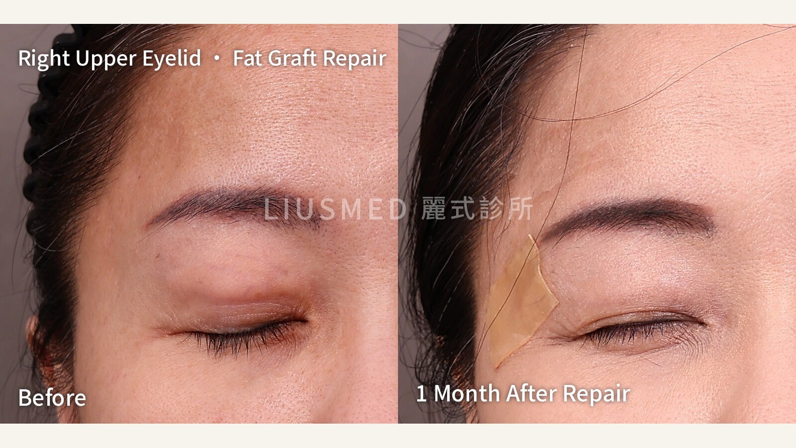
Fat grafted into the upper eyelids that now bulges too much can be removed. But upper-eyelid skin is thin, the lacrimal gland sits nearby and the eyeball lies beneath, so taking too much or too little both show on the surface. Our approach is to find out with ultrasound which layer the fat is in first, then remove it conservatively, in stages.
This is the record of one patient. She had only the right eye done first and wanted to see the result before deciding on the left. That gave the one-month photo a comparison that is hard to come by: same day, same light, same body weight, with the right eye treated and the left eye not yet treated.
Three months after the graft, the upper eyelids started to bulge
At the end of 2023 she had autologous fat grafted into the hollows of her upper eyelids, to soften a sunken look.
About three months later, the upper eyelids began to look clearly puffy: the whole lid was raised, with a visible contour line along its upper edge. After she gained around 5 kg, the bulge became more obvious. Grafted fat that survives can grow along with weight gain, and on skin as thin as the upper eyelid the change shows directly.
What ultrasound showed: the fat was placed superficially
On ultrasound, the fat sat in a relatively superficial layer.
Upper-eyelid skin is among the thinnest skin on the body. Fat placed a little deeper has muscle and tissue in front of it. Fat placed superficially has almost nothing to hide behind, so its outline shows even at rest.
It shows most when she squeezes her eyes shut. As the muscles around the eye contract, the superficial mass is pushed into a distinct shape:
The top row is before, taken in the procedure room on the day of surgery, so the lighting differs from the after photo. The bottom row is one month after. With the same squeeze, that mass in the right upper eyelid is no longer visible.
Why the upper eyelid is one of the more demanding areas for extraction
Among pinhole extractions, the upper eyelid is an advanced area, for three reasons:
- The skin is very thin. Take too much and a bulge becomes a hollow; take too little and it still shows. There is very little margin on the surface.
- The lacrimal gland sits at the outer upper corner. The extraction has to stay clear of it.
- The eyeball lies deeper in. The direction and depth of every instrument have to be tightly controlled.
So the upper eyelid is not a place for a "get it all out at once" mindset.
Staged extraction: conservative the first time
After the ultrasound assessment, my advice to her came down to three things:
- Confirm that this is superficially placed fat, and work at the layer where it sits.
- Remove it in stages. Take less the first time, wait until it has fully healed, then see whether anything still stands out, and only then consider a second session.
- Explain recovery before the procedure. There will be swelling and possibly bruising after extraction, and both fade over the recovery period.
The entry point is just outside the tail of the eyebrow. The tape on the temple in the after photo marks where it is; there is no opening on the eyelid itself. Anesthesia was local anesthesia plus gentle pain-relief anesthesia.
This is the tissue removed from the right upper eyelid that day:
The amount is small, and that is deliberate. The spot labelled L is empty because the left eye was not treated this time.
Only the right eye first: the comparison inside the same photo
She decided to have the right eye done first and to decide on the left after seeing the result.
That gives the one-month photo an extra layer of meaning. The usual questions about before-and-after photos are about the lighting, the angle, or whether the patient simply lost weight. In the bottom row of this photo, both eyes were taken on the same day, under the same light, at the same body weight, with the same camera:
The top row is before, with both upper eyelids bulging. The bottom row is one month after. The eye on the left side of the image is her right eye, which was treated; the eye on the right side of the image is her left eye, which has not been. The difference between the two shows what this extraction changed.
One month after the repair
At the one-month visit, the right upper eyelid was much smoother. We had left room for a second extraction, and after talking it through, no second session is planned for now.
Whether and when to treat the left eye is up to her, after she has seen how the right one turned out.
Why one month is not the end point
At one month, the tissue is still recovering. Residual swelling will go down and the tissue will settle over time; the final result settles at around three months.
That is the reason for staging the extraction in the first place: remove conservatively the first time, give the tissue time to show the result, then decide whether more is needed. Extraction is not about removing as much as possible. Take too much from an upper eyelid and a bulge turns into a hollow. Two things matter to me: removing the right amount, and leaving a smooth surface.
Photos are used with the patient's consent and show the same patient before and one month after the repair. This is an individual case and does not represent everyone's result. All four photo sets are also in the case gallery.
Common questions
My upper eyelids bulge after a fat graft. Can the fat be removed?
It can be assessed. Ultrasound comes first, to see which layer the fat is in and how far it spreads. Fat placed superficially shows its outline at rest and more clearly when the eyes are squeezed shut. Upper-eyelid extraction is usually staged, conservative the first time, with a second session considered after healing. Whether it can be removed, and in how many sessions, depends on individual assessment.
Can the upper eyelid look hollow after the fat is removed?
It can if too much is taken. Upper-eyelid skin is thin and the surface leaves very little margin, so it is better to take less the first time and see what still needs work after healing than to go all the way in one session.
Does it always take two sessions?
No. For this patient we had left room for a second session, but she was much smoother at one month and, after discussion, none is planned for now. Whether a second session is needed depends on whether anything still stands out after healing.
If only one side is treated, won't the two sides look different?
Until the other side is treated, the two sides will look different, which is exactly what this patient's photos show. Treating one side first was her own choice; the benefit is being able to see the result before deciding on the other side.
How long is the recovery?
Swelling and bruising are common after extraction and fade over the recovery period. The final result settles at around three months. Actual recovery time varies from person to person.
Where is the entry point? Is there a wound on the eyelid?
For this patient, the entry point was just outside the tail of the eyebrow, with no opening on the eyelid. The entry point is planned individually according to where the fat is.
If your upper eyelids have had a fat graft
If your upper eyelids were grafted with fat and now look fuller than expected, more so when you close your eyes tightly, or have become more noticeable after a change in weight, an ultrasound assessment is a good first step. Knowing which layer the fat is in and how far it spreads is what makes it possible to discuss whether, and how, to remove it.
For other reasons upper eyelids get puffier after filler or fat, see Sunken Upper Eyelid Filler or Fat Gone Wrong. How we handle the different fat graft complications is covered in fat graft complication repair.
You are welcome to describe your situation through our online consultation.
Further reading
- Sunken Upper Eyelid Filler or Fat Gone Wrong — Why It Looks Puffy or Droopy
- Fat Grafted Under the Eye Turned Into a Lump, Then a Stimulator Was Added to Hide It: A Full Record of Removing Five-Year Stacked Nodules
- Two Years of Ellansé Forehead Lumps: From Ultrasound Map to Three Months After Repair
About the author
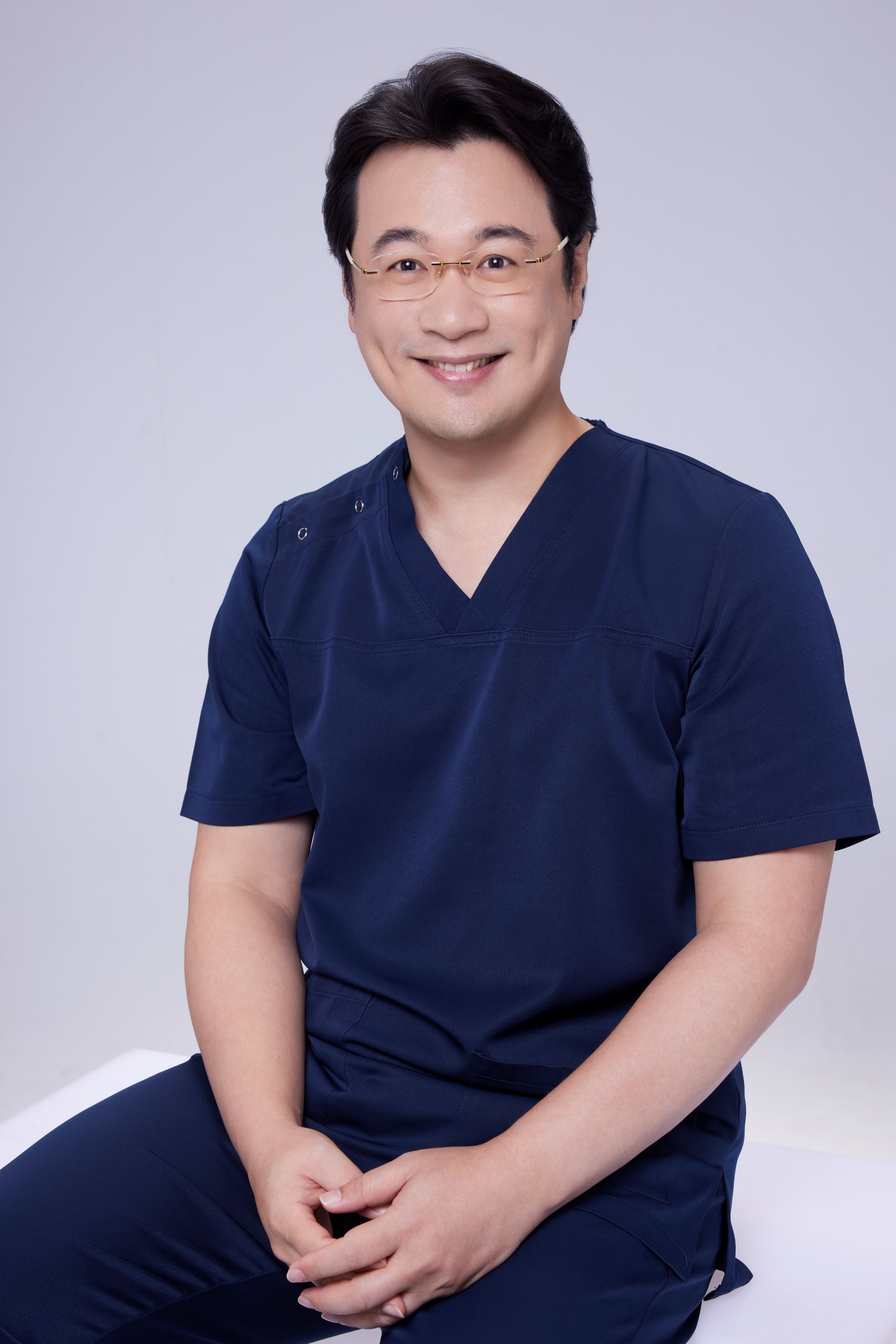
Dr. Ta-Ju Liu is the director of Liusmed Clinic. His work focuses on ultrasound-guided repair of filler complications, on the principle that you have to see it to handle it safely.
Related services
Specialties
Credentials
- Kaohsiung Medical University, School of Medicine
- Attending Physician, Dermatology, Kaohsiung Chang Gung Memorial Hospital
- Attending Physician, Aesthetic Center, Kaohsiung Chang Gung Memorial Hospital
- Visiting Physician, Dermatology, Xiamen Chang Gung Hospital
- Visiting Physician, Aesthetic Center, Xiamen Chang Gung Hospital
"For every surgery, I strive to achieve a good outcome through a small incision and refined technique. Minimally invasive surgery is not just a technique — it's a commitment of respect to every patient."
Recovery after any procedure needs peer support too


Want to learn more?
Schedule a consultation for professional evaluation and advice