Stone-Hard Lump at the Eye Corner After Fat Grafting, Removed Through One Pinhole
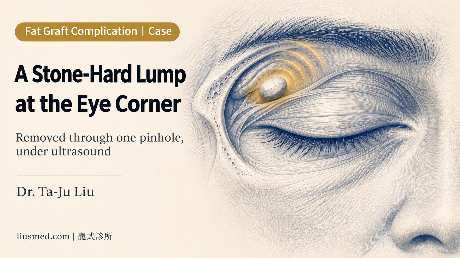
When I pressed on it, the lump slid away under my fingertip.
It was hard, as hard as a stone. It sat above the outer corner of her right eye, and with her eyes open you could barely see it from the outside. But a finger found it right away, and it moved under the finger.
Something this small is not easy to deal with. The skin of the upper eye socket is thin, the lacrimal gland (the tear gland) sits right beside it, and this lump was tucked inward. So we removed it bit by bit through a single pinhole, watching the ultrasound the whole time.
Fat grafting abroad a year ago, and a year later, this lump
A year ago she had autologous fat grafting (fat taken from her own body and injected) abroad, into the forehead and the eye sockets.
About a year later she felt a lump at the outer corner of her right eye. Just one. It was as hard as a stone, and it moved.
In the mirror you could hardly see it. Nothing showed on the outside, yet you could feel it with a finger.
The video: a record of this lump, just over a minute
We cut this case into a short video of just over a minute: pressing on the lump, the pre-procedure marking, how it looks with her eyes open, ultrasound before and after, the area pressed right afterward and the pinhole, and a same-angle comparison before and right after treatment. There is no narration; the explanations are on screen. The orbital anatomy illustration and the animation of the lump hardening are illustrative.
Barely visible from outside, clearly visible on ultrasound
With her eyes open, the skin at the outer corner looked ordinary. On ultrasound, there was a round dark area under the skin, the lump she had been feeling.
Ultrasound did two jobs here. First, it confirmed the lump was there and showed how big it was. Second, it showed how close it was to the skin and how far it pushed inward. Everything that came after depended on that map.
Key point: Not visible does not mean you can skip looking. At the outer eye corner, what your finger finds is only part of the picture; ultrasound shows the whole of it.
Why injected fat can slowly turn hard
Not all of the fat injected in a fat graft survives. The part that survives becomes part of the surrounding tissue. The part that does not has to be dealt with by the body.
Some of it is absorbed. Some of it gets walled off by a layer of tissue and, over time, slowly becomes fibrotic, changing from soft to hard. Hers was that kind: found about a year after the graft, and as hard as a stone.
For grafted fat that hardened under the eye instead, see the record of removing under-eye fat graft lumps. This article is only about this one lump at the outer eye corner.
Why this lump at the eye corner is harder to treat
The upper eye socket is not an easy area to begin with. The skin is thin, so there is very little room for error on the surface, and the lacrimal gland sits toward the upper outer side, so any treatment has to stay clear of it.
This lump added one more layer of difficulty. It lay in the skin layer, so it was shallow, but it bulged inward, and when pressed it felt about as deep as the bony orbital rim next to it. Under a fingertip, it was hard to tell where the lump ended and the edge of the bone began. And it moved.
| By touch | On ultrasound | |
|---|---|---|
| Can it be found? | Yes, but it moves and is hard to pin down | The whole mass is visible |
| Depth | Feels as deep as the orbital rim; hard to tell apart | Seen in the skin layer, bulging inward |
| During treatment | Relies on feel | Watched as it is done |
So for this lump, working by touch alone would not have been precise enough.
Key point: Some things are harder to treat the smaller they are. A small lump, thin skin, and the lacrimal gland next door leave very little room for error.
Watching the ultrasound, removing it bit by bit through one pinhole
The lump was located on ultrasound, and we went in through a single pinhole and removed it bit by bit while watching the screen. What comes out stays inside the needle, and the surface is left with one pinhole.
The skin at the eye corner is thin and the lacrimal gland is right there, so every step has to be visible on ultrasound before going further.
Right after: the dark mass is gone, and it is flat when pressed
When it was done, we looked again with ultrasound. The dark area was gone.
Right after the procedure, pressing on the spot where the lump had been, it was flat. On the surface there was one pinhole.
This is a record of the moment right after treatment. How the swelling settles and how long recovery takes vary from person to person, and for this patient I only have the immediate result, so this article does not describe anything later.
How this differs from the earlier upper eyelid case
I have written about another patient in Upper Eyelids Bulging After a Fat Graft. Both had problems after fat was grafted into the upper eye socket, and both were treated with ultrasound through a single pinhole. But the two situations were different.
In that case the fat had been placed superficially and in a larger amount, so the whole upper eyelid bulged, more so when she closed her eyes tightly, and it was removed in stages, conservatively. In this case it was a single, mobile, stone-hard lump that barely showed from the outside, and the difficulty was that it sat deep and could not be located reliably by touch.
The same area can produce very different problems. That is why we look first, every time, before deciding how to treat.
The video is shared with the patient's consent and shows the same patient before and right after treatment. This is an individual case and results vary. Bruising and swelling can occur after pinhole removal and recovery time differs from person to person; whether anything remains is checked again with ultrasound at follow-up.
Common questions
I feel a hard, mobile lump after fat grafting. What is it?
It may be grafted fat that did not survive, was walled off, and then became fibrotic and hardened. But a hard lump that moves can also be other things: migrated filler, a lymph node, or a lipoma can all feel similar. Ultrasound comes first, to see what it is and which layer it is in. See also Sliding Lump on Your Cheek: Lymph Node or Migrated Filler?
It cannot be seen from outside. Does it still need treatment?
Whether it shows and whether it should be treated are two separate questions. First confirm on ultrasound what it is, how big it is, and how it relates to the surrounding tissue, and then decide together whether and when to treat it.
Can a hardened fat lump be dissolved with an injection?
Hyaluronidase, the enzyme used to dissolve hyaluronic acid (HA) filler, has no effect on fat or hardened tissue. A steroid cannot tell the lump apart from the normal tissue around it, so the lump may not shrink while the surrounding area may sink first, and on skin as thin as the upper eye socket that shows directly. Lumps that have hardened like this usually need to be found and removed.
Why ultrasound? Can't it be removed by feel?
In this case the lump bulged inward, felt as deep as the orbital rim, and moved, so touch alone could not pin down its position. The lacrimal gland was also next to it. Watching the ultrasound is how the position can be pinned down.
Will it swell afterward? How long is recovery?
This record only covers the moment right after treatment: flat when pressed, with one pinhole. Swelling and recovery after that vary by individual and are explained at follow-up based on how things actually go.
Will it leave a scar?
Only one pinhole was used for this lump. The pinhole is small, but how a mark heals depends on skin type, location, and aftercare, and differs from person to person.
If you have had fat grafting and can feel a hard lump
If you have had fat grafted into your forehead, eye sockets, or around the eyes and can now feel a hard lump, whether or not it shows from the outside, an ultrasound assessment is a good first step. Knowing what it is, which layer it is in, and what lies next to it is what makes it possible to discuss whether and how to treat it.
The ways we handle hard lumps after fat grafting are gathered in fat graft complication repair.
You are welcome to describe your situation through our online consultation.
Further reading
- Upper Eyelids Bulging After a Fat Graft: Right Eye First, With the Comparison Inside the Same Photo
- Fat Grafted Under the Eye Turned Into a Lump, Then a Stimulator Was Added to Hide It: A Full Record of Removing Five-Year Stacked Nodules
- Sliding Lump on Your Cheek: Lymph Node or Migrated Filler? 3 Ways to Tell
About the author
Dr. Ta-Ju Liu is the director of Liusmed Clinic. His work focuses on ultrasound-guided repair of filler complications, on the principle that you have to see it to handle it safely.
Related services

Specialties
Credentials
- Kaohsiung Medical University, School of Medicine
- Attending Physician, Dermatology, Kaohsiung Chang Gung Memorial Hospital
- Attending Physician, Aesthetic Center, Kaohsiung Chang Gung Memorial Hospital
- Visiting Physician, Dermatology, Xiamen Chang Gung Hospital
- Visiting Physician, Aesthetic Center, Xiamen Chang Gung Hospital
"For every surgery, I strive to achieve a good outcome through a small incision and refined technique. Minimally invasive surgery is not just a technique — it's a commitment of respect to every patient."
Related Articles
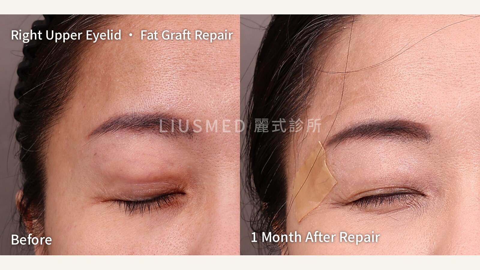
Upper Eyelids Bulging After a Fat Graft: Right Eye First, With the Comparison Inside the Same Photo
Fat was grafted into her upper eyelid hollows. Three months later the eyelids began to bulge, more so after weight gain. Ultrasound showed t
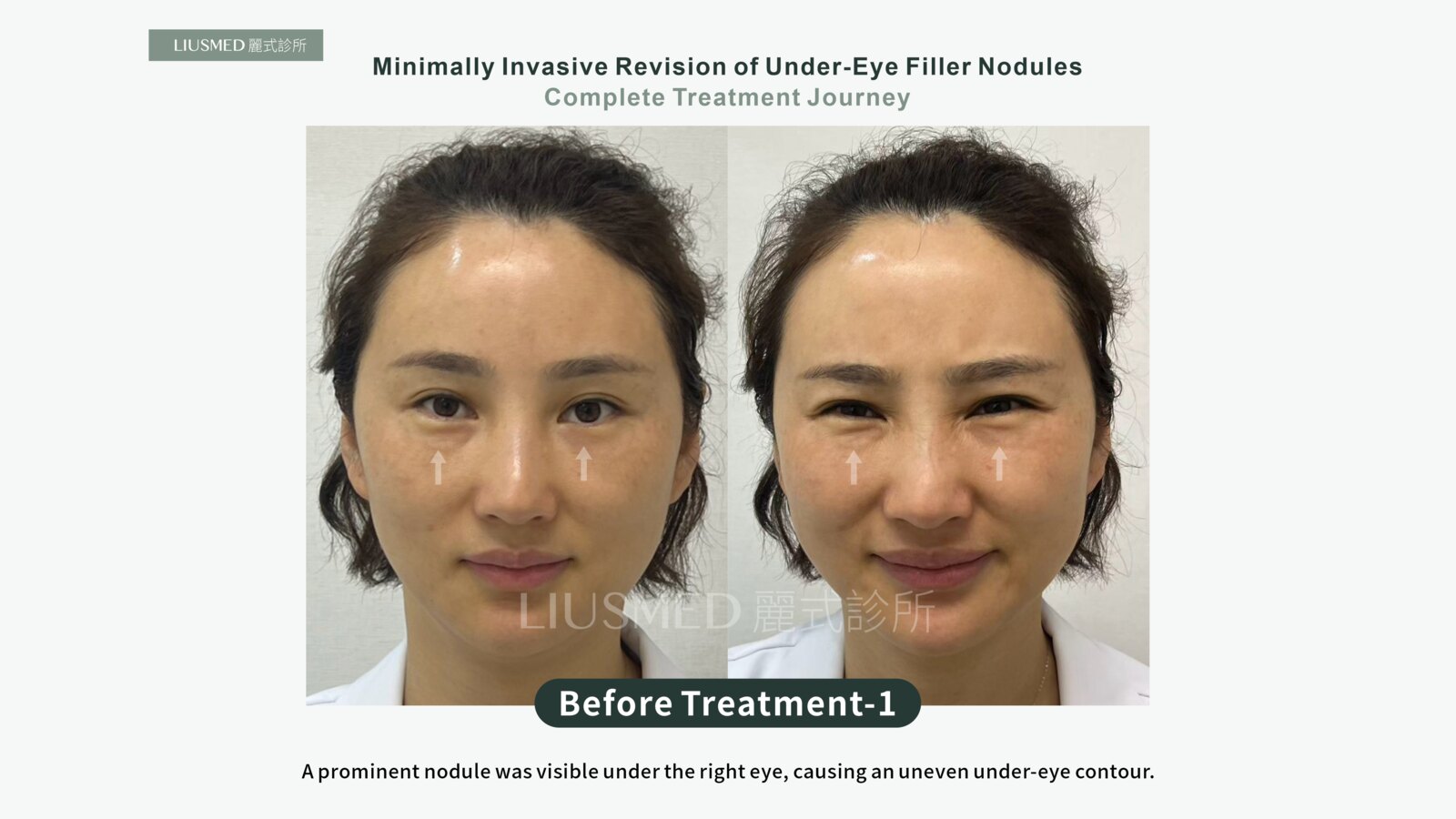
Fat Grafted Under the Eye Turned Into a Lump, Then a Stimulator Was Added to Hide It: A Full Record of Removing Five-Year Stacked Nodules
Fat was grafted under the eyes to fill the tear troughs, and it left a firm, visible bulge. To hide that bulge, a collagen stimulator was pl
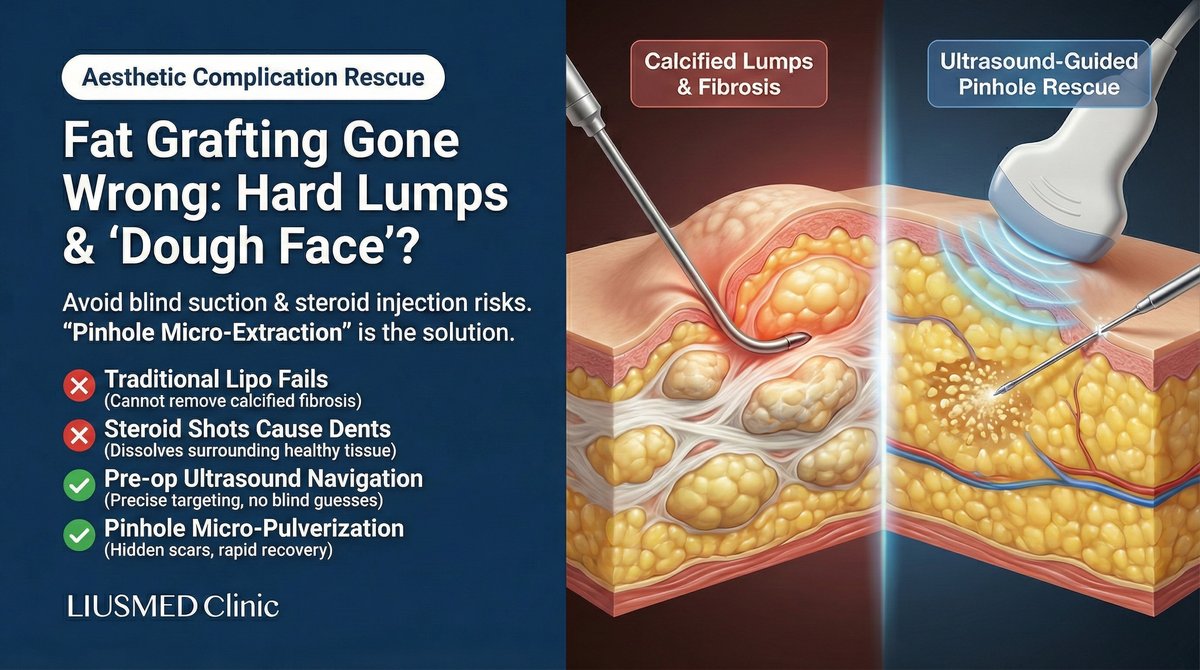
Fat Grafting Gone Wrong: FOS, Calcified Lumps & Granulomas
What to do when fat grafting goes wrong? Dr. Liu at Liusmed Clinic explains the pathology behind facial overfilling (FOS) and calcified lump
Recovery after any procedure needs peer support too


Want to learn more?
Schedule a consultation for professional evaluation and advice