Removing AlloDerm or a Dermis Graft Without Surgery: Shaving Only the Raised Part Through a Pinhole
In this article, "allogeneic dermis" means a sheet, strip or block of donor dermal scaffold (acellular dermal matrix, such as AlloDerm) placed directly under the skin. It is not the same thing as injectable dermal powder, and it differs in origin from an autologous dermis graft taken from your own body. The three fail in different ways, and they are removed in different ways.
Lately I have been seeing a new group of patients in consultation: people who had allogeneic or autologous dermis placed, and it did not settle well, leaving a ridge. Because the material went in as a whole strip or block, the problem shows up as a whole strip or block standing out, unlike injected dermal powder. These ridges can be removed.
This article is for you if you already know it is a dermis implant, you are thinking about having it taken out, and you may have been told that "dermis can never be removed cleanly." Whether to treat it and which options exist are covered in What to do about a dermis graft ridge. Here I only cover how it comes out: why it is hard, what open surgery costs, what ultrasound shows, and what it means to shave off only the raised part through a pinhole.
Key point: With a dermis implant, the question is not "can the whole strip be removed" but "how much, and from where." The goal is a smooth contour again, not an empty space where the graft used to be.
Why dermis is hard to remove: the body grows into it
Once a dermal scaffold is placed, the body gradually takes it in as its own tissue.
- In rats, host cells were already multiplying inside the scaffold seven days after implantation, with abundant early blood vessels (PMID 18349631).
- A rabbit study compared placement shapes. A single layer was essentially revascularized by day 14; rolled, thicker grafts were still not fully penetrated by day 28 (PMID 11304602).
- In human breast reconstruction, the scaffold was recolonized by fibroblasts and other host cells over the months after implantation, with blood vessels growing in (PMID 29569868).
- In ten adults with AlloDerm sheets placed under the skin behind the ear, biopsies over one year showed progressive repopulation of the graft by host cells (PMID 11368660).
Most of this comes from animals or breast surgery, so it cannot be mapped directly onto every face. But it all points the same way: dermis does not stay a "foreign block." It becomes part of the surrounding tissue.
What I see clinically is this: most dermis implants end up enclosed in a capsule, but a smaller share fuse tightly with the tissue around them.
An encapsulated graft is easier to remove surgically, but only if the skin is cut open. If the graft is being shaved through a single pinhole, whether it is tightly or loosely attached makes little difference to the difficulty. Neither is easy. The difficulty lies in the technique, not in the patient.
Three costs of cutting it out
The published literature describes dermis removal as an open procedure every time: the skin is opened, and the implant is lifted out with its capsule or scraped away. That is a reasonable surgical standard. On the face, though, it comes with three costs worth thinking through first.
First, the skin has to be opened. The implant is a whole strip, so the incision, and the scar it leaves, sits on the face, and is often not short.
Second, the small share that fused tightly is hard to scrape clean even when opened. Most encapsulated grafts come out whole; the tightly fused portion does not separate easily, even with direct access.
Third, once the whole strip is gone, the area tends to sink. The graft was placed to build that spot up. Take all of it out and nothing is left underneath, so a raised line becomes a sunken one.
Scarring and hollowing after facial excision are covered in more depth in Surgical excision: scarring and hollowing. Our clinic does not perform open removal; dermis implants are always handled through a pinhole.
What ultrasound can show
Ultrasound can see a dermis implant, and the literature uses it that way: a 14-patient study measured the height of autologous folded dermal grafts after rhinoplasty with ultrasound and found a 38% drop within six months (PMID 40728913).
Before removal, I use ultrasound to check:
- Which layer it sits in: how deep the graft lies under the skin and how far it is from the surface.
- How thick it is, and which segment rises above the contour: knowing the thickness tells us how much to take.
- How clear its edges are: whether there is a capsule, or whether it blends into the surrounding tissue.
- What is next to it: where the nearby vessels and nerves run, and whether there is fluid or inflammation.
Only with these answers can we decide where to shave, how much, and whether to stage it. How ultrasound guidance and single-pinhole extraction work is explained in How ultrasound-guided pinhole extraction works.
Pinhole shaving: controlling how much thickness comes out
Working through a pinhole of about 2 mm differs from surgery in one key way: we can control fairly precisely how much thickness of the graft is removed.
Say a strip was placed too thick. Shaving off 30% or 50% in one session can make the surface much smoother. The first session usually takes about 30 to 50%, to bring it level with the surrounding area rather than digging the whole strip out.
Surgery removes the whole strip; a pinhole can remove just the raised part. The two approaches solve different problems.
Clinics that treat dermis implants through a single ultrasound-guided pinhole are still uncommon in Taiwan. This approach grew out of our many years of removing fillers and implants.
Whether the graft is tightly or loosely attached, working through a pinhole is not easy. It does not always finish in one session, may need to be staged, and the result is not something we can promise in advance.
Whole-contour sculpting: taking out and putting in follow the same logic
This is how I think about every filler removal, and dermis happens to be a clear example of it.
When filler goes in, the aim is a balanced look, avoiding too much in one spot and overfilling. When something has been overfilled, it does not need to be removed 100%. Instead, following the natural lines of the face, the remaining filler is sculpted until it is smooth and even. Restoring natural lines and proper proportion is what gives a smooth, balanced result.
Cutting the whole strip open and removing it surgically tends to leave a local hollow at the same time. We can take out only the part that sticks out, so the line becomes smooth and natural.
"Surgery takes the whole strip out. Through a pinhole, we can shave off just the raised 30 to 50% so the surface sits level with its surroundings." — Dr. Ta-Ju Liu, October 2026
So how much to remove is decided by the lines and proportions of the whole face, not by "how much can come out." It looks a little different in each area:
- Nasolabial folds: the area I see most often. Typically a strip rises above the fold when smiling. See Nasolabial fold revision and Hard strip-like lumps in the nasolabial fold.
- Under-eye / aegyo-sal: next most common. The skin is very thin, so the edge of a horizontal strip is hard to hide. See Under-eye revision.
- Nasal dorsum: some people have it placed here. It shows as an extra ridge on the bridge, or a strip leaning to one side. See Nose revision.
- Depressed scars: a spot that used to be sunken ends up raised after grafting.
Is autologous dermis any different?
No. Autologous dermis (including dermis-fat grafts, which carry a layer of fat) comes from your own body, but once it is in place it is handled much like allogeneic dermis: ultrasound first to see position and thickness, then pinhole shaving of only the raised part.
The ultrasound study cited above used autologous folded dermis (PMID 40728913).
Recovery, staging and realistic expectations
The pinhole is small, but this is still a removal, so swelling and bruising will happen. We do not claim a short recovery.
| Item | Approximate time |
|---|---|
| Swelling and bruising settle | 2 to 3 weeks |
| Pinhole heals | About 2 weeks; usually hard to notice afterward, though this still depends on how your skin scars |
| Final result | About 3 months; varies by person |
| Interval between sessions, if staged | About 3 months |
Removing every last bit is not something we can promise. The goal is to improve it as much as is safely possible, bringing the contour back to natural proportions.
Common questions
Is an encapsulated dermis graft easier to remove?
For open surgery, yes. An encapsulated graft comes out whole more easily, but the skin has to be opened. Through a pinhole, a capsule or a tight attachment makes little difference. Neither is easy.
Why not remove all of it in one go?
Because the goal is a smooth line, not an empty space. The first session usually takes about 30 to 50%, which often smooths the surface a great deal; taking too much tends to leave a dent. If more is needed, we reassess after about three months.
How does the medical literature remove dermis grafts?
Every published description is an open procedure: the skin is opened and the implant is lifted out with its capsule or scraped away. Our clinic does not perform open removal; we always work under ultrasound guidance through a pinhole.
Do I need stitches afterward?
No. The pinhole is about 2 mm and heals in roughly two weeks. It is usually hard to notice afterward, although scarring still depends on your skin.
Is autologous dermis removed differently from allogeneic dermis?
Not really. Both are placed as a whole piece and both can be placed too thick. Both are checked with ultrasound first, and then only the raised part is shaved through a pinhole.
I had dermal powder injected. Is it treated the same way?
Not exactly. Dermal powder is injected as particles, and problems usually show up as a firm clump in one spot, which is closer to an ordinary filler lump. Start with Dermal powder hardening and fat-graft calcification.
Checking your own situation
If you had allogeneic or autologous dermis placed and can now feel a strip or see a block, the first step is an ultrasound to confirm which layer it sits in, how thick it is, and which segment rises above the contour. To talk through your situation, start with booking a consultation.
References
- Wong AK, Schonmeyr BH, Singh P, et al. Histologic analysis of angiogenesis and lymphangiogenesis in acellular human dermis. Plast Reconstr Surg. 2008;121(4):1144-1152. PMID: 18349631.
- Eppley BL. Experimental assessment of the revascularization of acellular human dermis for soft-tissue augmentation. Plast Reconstr Surg. 2001;107(3):757-762. PMID: 11304602.
- Boháč M, Danišovič Ľ, Koller J, et al. What happens to an acellular dermal matrix after implantation in the human body? A histological and electron microscopic study. Eur J Histochem. 2018;62(1):2873. PMID: 29569868.
- Sclafani AP, Romo T 3rd, Jacono AA, et al. Evaluation of acellular dermal graft (AlloDerm) sheet for soft tissue augmentation: a 1-year follow-up of clinical observations and histological findings. Arch Facial Plast Surg. 2001;3(2):101-103. PMID: 11368660.
- Kim DG, Kim KH, Cha HG, et al. Volume Retention of Folded Dermal Graft After Augmentation Rhinoplasty. J Craniofac Surg. 2025. doi:10.1097/SCS.0000000000011723. PMID: 40728913.
Related services
Medical review note: This article was written by Dr. Ta-Ju Liu based on clinical experience and the current literature. It is for education only and does not replace an in-person consultation. The open removal described here is the approach reported in the literature; our clinic uses ultrasound-guided, single-pinhole removal. Whether it suits you, and how it would be done, is decided only after an in-person examination and ultrasound assessment.
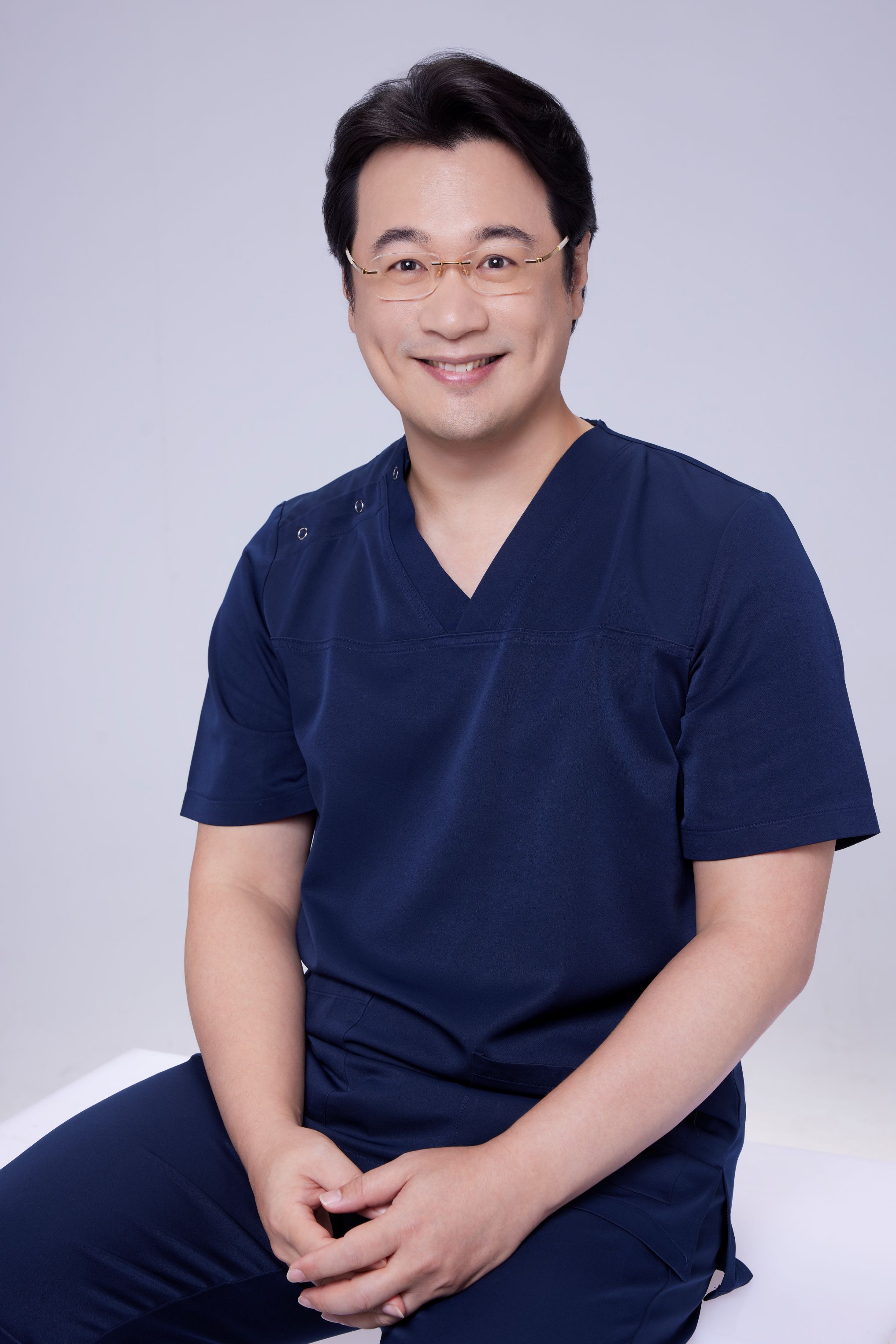
Specialties
Credentials
- Kaohsiung Medical University, School of Medicine
- Attending Physician, Dermatology, Kaohsiung Chang Gung Memorial Hospital
- Attending Physician, Aesthetic Center, Kaohsiung Chang Gung Memorial Hospital
- Visiting Physician, Dermatology, Xiamen Chang Gung Hospital
- Visiting Physician, Aesthetic Center, Xiamen Chang Gung Hospital
"For every surgery, I strive to achieve a good outcome through a small incision and refined technique. Minimally invasive surgery is not just a technique — it's a commitment of respect to every patient."
Recovery after any procedure needs peer support too


Want to learn more?
Schedule a consultation for professional evaluation and advice